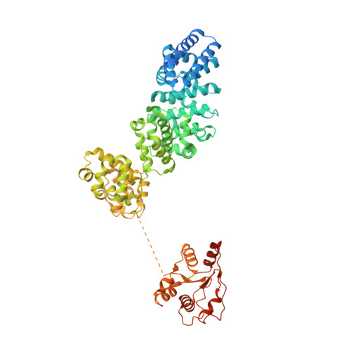
7CM7

NAD+-bound Sarm1 E642A in the self-inhibited state


Experimental Data Snapshot

  • Method: ELECTRON MICROSCOPY
  • Resolution: 2.60 Å
  • Aggregation State: PARTICLE 
  • Reconstruction Method: SINGLE PARTICLE 

wwPDB Validation   3D Report Full Report


This is version 1.3 of the entry. See complete history


Literature

The NAD + -mediated self-inhibition mechanism of pro-neurodegenerative SARM1.

Jiang, Y.Liu, T.Lee, C.H.Chang, Q.Yang, J.Zhang, Z.

(2020) Nature 588: 658-663

  • DOI: https://doi.org/10.1038/s41586-020-2862-z
  • Primary Citation of Related Structures:  
    7CM5, 7CM6, 7CM7

  • PubMed Abstract: 

    Pathological degeneration of axons disrupts neural circuits and represents one of the hallmarks of neurodegeneration 1-4 . Sterile alpha and Toll/interleukin-1 receptor motif-containing protein 1 (SARM1) is a central regulator of this neurodegenerative process 5-8 , and its Toll/interleukin-1 receptor (TIR) domain exerts its pro-neurodegenerative action through NADase activity 9,10 . However, the mechanisms by which the activation of SARM1 is stringently controlled are unclear. Here we report the cryo-electron microscopy structures of full-length SARM1 proteins. We show that NAD + is an unexpected ligand of the armadillo/heat repeat motifs (ARM) domain of SARM1. This binding of NAD + to the ARM domain facilitated the inhibition of the TIR-domain NADase through the domain interface. Disruption of the NAD + -binding site or the ARM-TIR interaction caused constitutive activation of SARM1 and thereby led to axonal degeneration. These findings suggest that NAD + mediates self-inhibition of this central pro-neurodegenerative protein.


  • Organizational Affiliation

    State Key Laboratory of Membrane Biology, Center for Life Sciences, School of Life Sciences, Peking University, Beijing, China.


Macromolecules
Find similar proteins by:  (by identity cutoff)  |  3D Structure
Entity ID: 1
MoleculeChains Sequence LengthOrganismDetailsImage
NAD(+) hydrolase SARM1
A, B, C, D, E
A, B, C, D, E, F, G, H
733Homo sapiensMutation(s): 1 
Gene Names: SARM1KIAA0524SAMD2SARM
EC: 3.2.2.6 (PDB Primary Data), 3.2.2 (PDB Primary Data)
UniProt & NIH Common Fund Data Resources
Find proteins for Q6SZW1 (Homo sapiens)
Explore Q6SZW1 
Go to UniProtKB:  Q6SZW1
PHAROS:  Q6SZW1
GTEx:  ENSG00000004139 
Entity Groups  
Sequence Clusters30% Identity50% Identity70% Identity90% Identity95% Identity100% Identity
UniProt GroupQ6SZW1
Sequence Annotations
Expand
  • Reference Sequence
Small Molecules
Ligands 1 Unique
IDChains Name / Formula / InChI Key2D Diagram3D Interactions
NAD (Subject of Investigation/LOI)
Query on NAD

Download Ideal Coordinates CCD File 
I [auth A]
J [auth B]
K [auth C]
L [auth D]
M [auth E]
I [auth A],
J [auth B],
K [auth C],
L [auth D],
M [auth E],
N [auth F],
O [auth G],
P [auth H]
NICOTINAMIDE-ADENINE-DINUCLEOTIDE
C21 H27 N7 O14 P2
BAWFJGJZGIEFAR-NNYOXOHSSA-N
Experimental Data & Validation

Experimental Data

  • Method: ELECTRON MICROSCOPY
  • Resolution: 2.60 Å
  • Aggregation State: PARTICLE 
  • Reconstruction Method: SINGLE PARTICLE 
EM Software:
TaskSoftware PackageVersion
RECONSTRUCTIONRELION

Structure Validation

View Full Validation Report


Entry History 

Deposition Data

Revision History  (Full details and data files)

  • Version 1.0: 2020-10-21
    Type: Initial release
  • Version 1.1: 2020-11-04
    Changes: Database references
  • Version 1.2: 2021-01-13
    Changes: Database references
  • Version 1.3: 2024-03-27
    Changes: Data collection, Database references