7CJA | pdb_00007cja

Crystal structure of TTK kinase domain in complex with compound 28


Experimental Data Snapshot

  • Method: X-RAY DIFFRACTION
  • Resolution: 2.49 Å
  • R-Value Free: 
    0.253 (Depositor), 0.250 (DCC) 
  • R-Value Work: 
    0.207 (Depositor), 0.210 (DCC) 
  • R-Value Observed: 
    0.212 (Depositor) 

Starting Model: experimental
View more details

wwPDB Validation   3D Report Full Report


Ligand Structure Quality Assessment 

Created with Raphaël 2.3.0Worse 01 BetterLigand structure goodness of fit to experimental dataBest fitted G0UClick on this verticalbar to view details

This is version 1.3 of the entry. See complete history


Literature

X-ray Crystal Structure-Guided Design and Optimization of 7 H -Pyrrolo[2,3- d ]pyrimidine-5-carbonitrile Scaffold as a Potent and Orally Active Monopolar Spindle 1 Inhibitor.

Lee, Y.Kim, H.Kim, H.Cho, H.Y.Jee, J.G.Seo, K.A.Son, J.B.Ko, E.Choi, H.G.Kim, N.D.Kim, I.

(2021) J Med Chem 64: 6985-6995

  • DOI: https://doi.org/10.1021/acs.jmedchem.1c00542
  • Primary Citation of Related Structures:  
    7CHM, 7CHN, 7CHT, 7CIL, 7CJA, 7CLH

  • PubMed Abstract: 

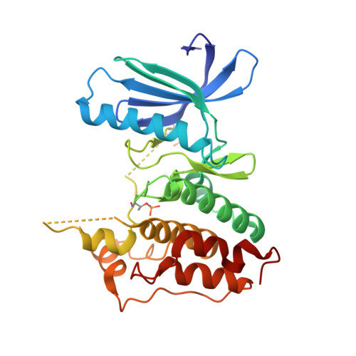
    Triple-negative breast cancer (TNBC) is an aggressive breast-cancer subtype associated with poor prognosis and high relapse rates. Monopolar spindle 1 kinase (MPS1) is an apical dual-specificity protein kinase that is over-expressed in TNBC. We herein report a highly selective MPS1 inhibitor based on a 7 H -pyrrolo[2,3- d ]pyrimidine-5-carbonitrile scaffold. Our lead optimization was guided by key X-ray crystal structure analysis. In vivo evaluation of candidate ( 9 ) is shown to effectively mitigate human TNBC cell proliferation.


  • Organizational Affiliation

    College of Pharmacy and Yonsei Institute of Pharmaceutical Sciences, Yonsei University, 85 Songdogwahak-ro, Yeonsu-gu, Incheon 21983, South Korea.


Macromolecules
Find similar proteins by:  (by identity cutoff)  |  3D Structure
Entity ID: 1
MoleculeChains Sequence LengthOrganismDetailsImage
Dual specificity protein kinase TTK283Homo sapiensMutation(s): 0 
Gene Names: TTKMPS1MPS1L1
EC: 2.7.12.1
UniProt & NIH Common Fund Data Resources
Find proteins for P33981 (Homo sapiens)
Explore P33981 
Go to UniProtKB:  P33981
PHAROS:  P33981
GTEx:  ENSG00000112742 
Entity Groups  
Sequence Clusters30% Identity50% Identity70% Identity90% Identity95% Identity100% Identity
UniProt GroupP33981
Sequence Annotations
Expand
  • Reference Sequence
Small Molecules
Ligands 1 Unique
IDChains Name / Formula / InChI Key2D Diagram3D Interactions
G0U (Subject of Investigation/LOI)
Query on G0U

Download Ideal Coordinates CCD File 
B [auth A]4-(cyclopentylmethylamino)-2-[[2-methoxy-4-(2-oxidanylidenepyrrolidin-1-yl)phenyl]amino]-7H-pyrrolo[2,3-d]pyrimidine-5-carbonitrile
C24 H27 N7 O2
HNTNJDLDWXMVRF-UHFFFAOYSA-N
Modified Residues  2 Unique
IDChains TypeFormula2D DiagramParent
SEP
Query on SEP
A
L-PEPTIDE LINKINGC3 H8 N O6 PSER
TPO
Query on TPO
A
L-PEPTIDE LINKINGC4 H10 N O6 PTHR
Experimental Data & Validation

Experimental Data

  • Method: X-RAY DIFFRACTION
  • Resolution: 2.49 Å
  • R-Value Free:  0.253 (Depositor), 0.250 (DCC) 
  • R-Value Work:  0.207 (Depositor), 0.210 (DCC) 
  • R-Value Observed: 0.212 (Depositor) 
Space Group: I 2 2 2
Unit Cell:
Length ( Å )Angle ( ˚ )
a = 70.644α = 90
b = 110.536β = 90
c = 112.456γ = 90
Software Package:
Software NamePurpose
PHENIXrefinement
HKL-2000data reduction
HKL-2000data scaling
PHENIXphasing

Structure Validation

View Full Validation Report


Ligand Structure Quality Assessment 

Created with Raphaël 2.3.0Worse 01 BetterLigand structure goodness of fit to experimental dataBest fitted G0UClick on this verticalbar to view details

Entry History 

Deposition Data

Revision History  (Full details and data files)

  • Version 1.0: 2021-05-12
    Type: Initial release
  • Version 1.1: 2021-06-09
    Changes: Database references
  • Version 1.2: 2023-11-29
    Changes: Data collection, Database references, Refinement description
  • Version 1.3: 2024-11-13
    Changes: Structure summary