7CIY | pdb_00007ciy

Crystal structure of N191G-mutated tyrosinase from Streptomyces castaneoglobisporus in complex with the caddie protein obtained by soaking in the solution containing Cu(II) and hydroxylamine for 24 h


Experimental Data Snapshot

  • Method: X-RAY DIFFRACTION
  • Resolution: 1.47 Å
  • R-Value Free: 
    0.191 (Depositor), 0.173 (DCC) 
  • R-Value Work: 
    0.137 (Depositor) 
  • R-Value Observed: 
    0.137 (Depositor) 

Starting Model: experimental
View more details

wwPDB Validation 3D Report Full Report

Validation slider image for 7CIY

This is version 1.2 of the entry. See complete history

Literature

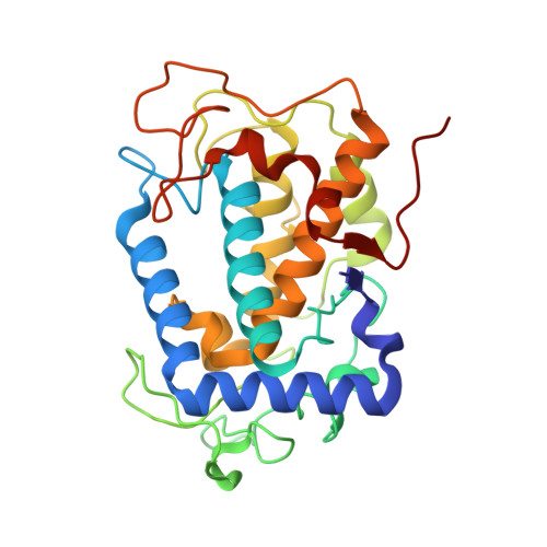
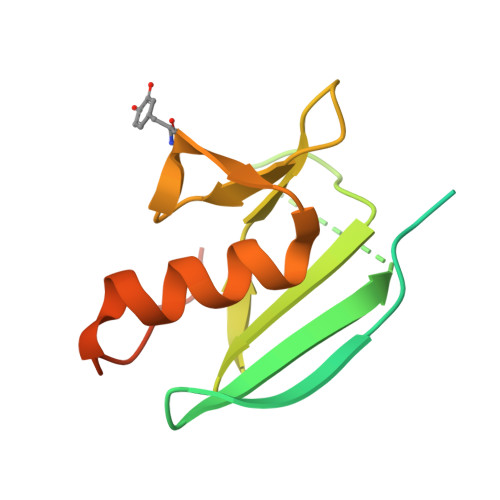
The basicity of an active-site water molecule discriminates between tyrosinase and catechol oxidase activity.

Matoba, Y.Oda, K.Muraki, Y.Masuda, T.

(2021) Int J Biol Macromol 183: 1861-1870

  • DOI: https://doi.org/10.1016/j.ijbiomac.2021.05.206
  • Primary Citation Related Structures: 
    7CIT, 7CIY

  • PubMed Abstract: 

    Tyrosinase (Ty) and catechol oxidase (CO) are members of type-3 copper enzymes. While Ty catalyzes both phenolase and catecholase reactions, CO catalyzes only the latter reaction. In the present study, Ty was found to catalyze the catecholase reaction, but hardly the phenolase reaction in the presence of the metallochaperon called "caddie protein (Cad)". The ability of the substrates to dissociate the motif shielding the active-site pocket seems to contribute critically to the substrate specificity of Ty. In addition, a mutation at the N191 residue, which forms a hydrogen bond with a water molecule near the active center, decreased the inherent ratio of phenolase versus catecholase activity. Unlike the wild-type complex, reaction intermediates were not observed when the catalytic reaction toward the Y98 residue of Cad was progressed in the crystalline state. The increased basicity of the water molecule may be necessary to inhibit the proton transfer from the conjugate acid to a hydroxide ion bridging the two copper ions. The deprotonation of the substrate hydroxyl by the bridging hydroxide seems to be significant for the efficient catalytic cycle of the phenolase reaction.


  • Organizational Affiliation
    • Faculty of Pharmacy, Yasuda Women's University, Yasuhigashi 6-13-1, Asaminami-ku, Hiroshima, 731-0153, Japan. Electronic address: matoba@yasuda-u.ac.jp.

Macromolecule Content 

  • Total Structure Weight: 46.85 kDa 
  • Atom Count: 3,279 
  • Modeled Residue Count: 356 
  • Deposited Residue Count: 415 
  • Unique protein chains: 2

Macromolecules

Find similar proteins by:|  3D Structure
Entity ID: 1
MoleculeChains  Sequence LengthOrganismDetailsImage
Tyrosinase281Streptomyces castaneoglobisporusMutation(s): 0 
UniProt
Find proteins for Q83WS2 (Streptomyces castaneoglobisporus)
Explore Q83WS2 
Go to UniProtKB:  Q83WS2
Entity Groups
Sequence Clusters30% Identity50% Identity70% Identity90% Identity95% Identity100% Identity
UniProt GroupQ83WS2
Sequence Annotations
Expand
Reference Sequence
Find similar proteins by:|  3D Structure
Entity ID: 2
MoleculeChains  Sequence LengthOrganismDetailsImage
MelC134Streptomyces castaneoglobisporusMutation(s): 0 
UniProt
Find proteins for Q83WS1 (Streptomyces castaneoglobisporus)
Explore Q83WS1 
Go to UniProtKB:  Q83WS1
Entity Groups
Sequence Clusters30% Identity50% Identity70% Identity90% Identity95% Identity100% Identity
UniProt GroupQ83WS1
Sequence Annotations
Expand
Reference Sequence

Small Molecules

Ligands 3 Unique
IDChains Name / Formula / InChI Key2D Diagram3D Interactions
CU
(Subject of Investigation/LOI)

Query on CU


Download:Ideal Coordinates CCD File
D [auth A],
E [auth A],
F [auth A],
J [auth B]
COPPER (II) ION
Cu
JPVYNHNXODAKFH-UHFFFAOYSA-N
NO3

Query on NO3


Download:Ideal Coordinates CCD File
G [auth A],
H [auth A],
I [auth A],
K [auth B],
L [auth B]
NITRATE ION
N O3
NHNBFGGVMKEFGY-UHFFFAOYSA-N
PEO

Query on PEO


Download:Ideal Coordinates CCD File
C [auth A]HYDROGEN PEROXIDE
H2 O2
MHAJPDPJQMAIIY-UHFFFAOYSA-N
Modified Residues  1 Unique
IDChains TypeFormula2D DiagramParent
G1X
Query on G1X
B
L-PEPTIDE LINKINGC9 H9 N O4TYR

Experimental Data & Validation

Experimental Data

  • Method: X-RAY DIFFRACTION
  • Resolution: 1.47 Å
  • R-Value Free:  0.191 (Depositor), 0.173 (DCC) 
  • R-Value Work:  0.137 (Depositor) 
  • R-Value Observed: 0.137 (Depositor) 
Space Group: P 21 21 2
Unit Cell:
Length ( Å )Angle ( ˚ )
a = 64.85α = 90
b = 97.4β = 90
c = 54.98γ = 90
Software Package:
Software NamePurpose
HKL-2000data scaling
SHELXrefinement
PDB_EXTRACTdata extraction
HKL-2000data reduction
MOLREPphasing

Structure Validation

View Full Validation Report


Entry History 

Deposition Data

Revision History  (Full details and data files)

  • Version 1.0: 2021-06-16
    Type: Initial release
  • Version 1.1: 2021-06-23
    Changes: Database references
  • Version 1.2: 2023-11-29
    Changes: Data collection, Database references, Derived calculations, Refinement description