7CIH | pdb_00007cih

Microbial Hormone-sensitive lipase E53 mutant S285G


Experimental Data Snapshot

  • Method: X-RAY DIFFRACTION
  • Resolution: 1.79 Å
  • R-Value Free: 
    0.190 (Depositor), 0.189 (DCC) 
  • R-Value Work: 
    0.169 (Depositor) 

Starting Model: experimental
View more details

wwPDB Validation 3D Report Full Report

Validation slider image for 7CIH

Ligand Structure Quality Assessment 


This is version 1.2 of the entry. See complete history

Literature

Mechanism and Structural Insights Into a Novel Esterase, E53, Isolated From Erythrobacter longus .

Ding, Y.Nie, L.Yang, X.C.Li, Y.Huo, Y.Y.Li, Z.Gao, Y.Cui, H.L.Li, J.Xu, X.W.

(2021) Front Microbiol 12: 798194-798194

  • DOI: https://doi.org/10.3389/fmicb.2021.798194
  • Primary Citation Related Structures: 
    7CI0, 7CIH, 7W8N

  • PubMed Abstract: 

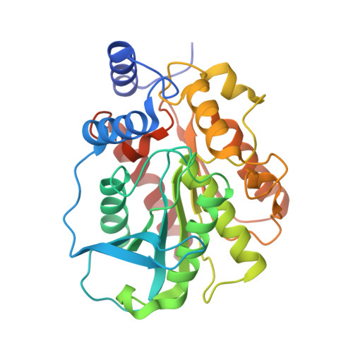
    Esterases are a class of enzymes that split esters into an acid and an alcohol in a chemical reaction with water, having high potential in pharmaceutical, food and biofuel industrial applications. To advance the understanding of esterases, we have identified and characterized E53, an alkalophilic esterase from a marine bacterium Erythrobacter longus . The crystal structures of wild type E53 and three variants were solved successfully using the X-ray diffraction method. Phylogenetic analysis classified E53 as a member of the family IV esterase. The enzyme showed highest activity against p -nitrophenyl butyrate substrate at pH 8.5-9.5 and 40 ° C. Based on the structural feature, the catalytic pocket was defined as R1 (catalytic center), R2 (pocket entrance), and R3 (end area of pocket) regions. Nine variants were generated spanning R1-R3 and thorough functional studies were performed. Detailed structural analysis and the results obtained from the mutagenesis study revealed that mutations in the R1 region could regulate the catalytic reaction in both positive and negative directions; expanding the bottleneck in R2 region has improved the enzymatic activity; and R3 region was associated with the determination of the pH pattern of E53. N166A in R3 region showed reduced activity only under alkaline conditions, and structural analysis indicated the role of N166 in stabilizing the loop by forming a hydrogen bond with L193 and G233. In summary, the systematic studies on E53 performed in this work provide structural and functional insights into alkaliphilic esterases and further our knowledge of these enzymes.


  • Organizational Affiliation
    • Key Laboratory of Marine Ecosystem Dynamics, Second Institute of Oceanography, Ministry of Natural Resources, Hangzhou, China.

Macromolecule Content 

  • Total Structure Weight: 137.34 kDa 
  • Atom Count: 10,685 
  • Modeled Residue Count: 1,240 
  • Deposited Residue Count: 1,256 
  • Unique protein chains: 1

Macromolecules

Find similar proteins by:|  3D Structure
Entity ID: 1
MoleculeChains  Sequence LengthOrganismDetailsImage
Lipase
A, B, C, D
314Erythrobacter longusMutation(s): 1 
Gene Names: EH31_02760
UniProt
Find proteins for A0A074MDU6 (Erythrobacter longus)
Explore A0A074MDU6 
Go to UniProtKB:  A0A074MDU6
Entity Groups
Sequence Clusters30% Identity50% Identity70% Identity90% Identity95% Identity100% Identity
UniProt GroupA0A074MDU6
Sequence Annotations
Expand
Reference Sequence

Small Molecules

Ligands 7 Unique
IDChains Name / Formula / InChI Key2D Diagram3D Interactions
D8F
(Subject of Investigation/LOI)

Query on D8F


Download:Ideal Coordinates CCD File
E [auth A],
JA [auth C],
S [auth B],
TA [auth D]
(4-nitrophenyl) hexanoate
C12 H15 N O4
OLRXUEYZKCCEKK-UHFFFAOYSA-N
NPO

Query on NPO


Download:Ideal Coordinates CCD File
DA [auth B],
EA [auth B]
P-NITROPHENOL
C6 H5 N O3
BTJIUGUIPKRLHP-UHFFFAOYSA-N
SO4

Query on SO4


Download:Ideal Coordinates CCD File
EB [auth D],
HA [auth B]
SULFATE ION
O4 S
QAOWNCQODCNURD-UHFFFAOYSA-L
GOL

Query on GOL


Download:Ideal Coordinates CCD File
G [auth A]
I [auth A]
KA [auth C]
LA [auth C]
MA [auth C]
G [auth A],
I [auth A],
KA [auth C],
LA [auth C],
MA [auth C],
NA [auth C],
R [auth B],
SA [auth C],
UA [auth D],
X [auth B]
GLYCEROL
C3 H8 O3
PEDCQBHIVMGVHV-UHFFFAOYSA-N
DIO

Query on DIO


Download:Ideal Coordinates CCD File
BB [auth D],
VA [auth D],
W [auth B],
WA [auth D]
1,4-DIETHYLENE DIOXIDE
C4 H8 O2
RYHBNJHYFVUHQT-UHFFFAOYSA-N
DMS

Query on DMS


Download:Ideal Coordinates CCD File
CA [auth B]
CB [auth D]
DB [auth D]
F [auth A]
GA [auth B]
CA [auth B],
CB [auth D],
DB [auth D],
F [auth A],
GA [auth B],
IA [auth C],
J [auth A],
K [auth A],
P [auth A],
Q [auth A],
RA [auth C],
U [auth B],
XA [auth D]
DIMETHYL SULFOXIDE
C2 H6 O S
IAZDPXIOMUYVGZ-UHFFFAOYSA-N
EDO

Query on EDO


Download:Ideal Coordinates CCD File
AA [auth B]
AB [auth D]
BA [auth B]
FA [auth B]
H [auth A]
AA [auth B],
AB [auth D],
BA [auth B],
FA [auth B],
H [auth A],
L [auth A],
M [auth A],
N [auth A],
O [auth A],
OA [auth C],
PA [auth C],
QA [auth C],
T [auth B],
V [auth B],
Y [auth B],
YA [auth D],
Z [auth B],
ZA [auth D]
1,2-ETHANEDIOL
C2 H6 O2
LYCAIKOWRPUZTN-UHFFFAOYSA-N

Experimental Data & Validation

Experimental Data

  • Method: X-RAY DIFFRACTION
  • Resolution: 1.79 Å
  • R-Value Free:  0.190 (Depositor), 0.189 (DCC) 
  • R-Value Work:  0.169 (Depositor) 
Space Group: P 21 2 21
Unit Cell:
Length ( Å )Angle ( ˚ )
a = 70.664α = 90
b = 129.692β = 90
c = 219.567γ = 90
Software Package:
Software NamePurpose
REFMACrefinement
XDSdata reduction
XDSdata scaling
PHENIXphasing

Structure Validation

View Full Validation Report


Ligand Structure Quality Assessment 


Entry History 

& Funding Information

Deposition Data


Funding OrganizationLocationGrant Number
National Science Foundation (NSF, China)China31770004

Revision History  (Full details and data files)

  • Version 1.0: 2021-07-14
    Type: Initial release
  • Version 1.1: 2022-02-23
    Changes: Database references
  • Version 1.2: 2023-11-29
    Changes: Data collection, Refinement description