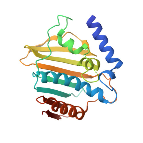
7C7N

Crystal structure of E.coli DNA gyrase B in complex with 6-fluoro-8-(methylamino)-2-oxo-1,2-dihydroquinoline derivative


Experimental Data Snapshot

  • Method: X-RAY DIFFRACTION
  • Resolution: 2.30 Å
  • R-Value Free: 0.276 
  • R-Value Work: 0.206 
  • R-Value Observed: 0.209 

Starting Model: experimental
View more details

wwPDB Validation   3D Report Full Report


Ligand Structure Quality Assessment 


This is version 1.2 of the entry. See complete history


Literature

Lead optimization of 8-(methylamino)-2-oxo-1,2-dihydroquinolines as bacterial type II topoisomerase inhibitors.

Ushiyama, F.Amada, H.Mihara, Y.Takeuchi, T.Tanaka-Yamamoto, N.Mima, M.Kamitani, M.Wada, R.Tamura, Y.Endo, M.Masuko, A.Takata, I.Hitaka, K.Sugiyama, H.Ohtake, N.

(2020) Bioorg Med Chem 28: 115776-115776

  • DOI: https://doi.org/10.1016/j.bmc.2020.115776
  • Primary Citation of Related Structures:  
    7C7N, 7C7O

  • PubMed Abstract: 

    The global increase in multidrug-resistant pathogens has caused severe problems in the treatment of infections. To overcome these difficulties, the advent of a new chemical class of antibacterial drug is eagerly desired. We aimed at creating novel antibacterial agents against bacterial type II topoisomerases, which are well-validated targets. TP0480066 (compound 32) has been identified by using structure-based optimization originated from lead compound 1, which was obtained as a result of our previous lead identification studies. The MIC 90 values of TP0480066 against methicillin-resistant Staphylococcus aureus (MRSA), vancomycin-resistant Enterococci (VRE), and genotype penicillin-resistant Streptococcus pneumoniae (gPRSP) were 0.25, 0.015, and 0.06 μg/mL, respectively. Hence, TP0480066 can be regarded as a promising antibacterial drug candidate of this chemical class.


  • Organizational Affiliation

    Taisho Pharmaceutical Co., Ltd, 1-403 Yoshino-Cho, Kita-Ku, Saitama 331-9530, Japan. Electronic address: f-ushiyama@taisho.co.jp.


Macromolecules
Find similar proteins by:  (by identity cutoff)  |  3D Structure
Entity ID: 1
MoleculeChains Sequence LengthOrganismDetailsImage
DNA gyrase subunit B220Escherichia coliMutation(s): 0 
Gene Names: gyrBNCTC10766_01911NCTC9007_05029
EC: 5.6.2.2
UniProt
Find proteins for P0AES6 (Escherichia coli (strain K12))
Explore P0AES6 
Go to UniProtKB:  P0AES6
Entity Groups  
Sequence Clusters30% Identity50% Identity70% Identity90% Identity95% Identity100% Identity
UniProt GroupP0AES6
Sequence Annotations
Expand
  • Reference Sequence
Small Molecules
Ligands 1 Unique
IDChains Name / Formula / InChI Key2D Diagram3D Interactions
FKR (Subject of Investigation/LOI)
Query on FKR

Download Ideal Coordinates CCD File 
B [auth A]4-[[6-fluoranyl-8-(methylamino)-2-oxidanylidene-1~{H}-quinolin-3-yl]carbonylamino]benzoic acid
C18 H14 F N3 O4
DFAPUBLMKMWKJW-UHFFFAOYSA-N
Experimental Data & Validation

Experimental Data

  • Method: X-RAY DIFFRACTION
  • Resolution: 2.30 Å
  • R-Value Free: 0.276 
  • R-Value Work: 0.206 
  • R-Value Observed: 0.209 
  • Space Group: P 21 21 21
Unit Cell:
Length ( Å )Angle ( ˚ )
a = 41.333α = 90
b = 67.052β = 90
c = 68.213γ = 90
Software Package:
Software NamePurpose
REFMACrefinement
CrystalCleardata reduction
CrystalCleardata scaling
MOLREPphasing

Structure Validation

View Full Validation Report


Ligand Structure Quality Assessment 


Entry History 

Deposition Data

Revision History  (Full details and data files)

  • Version 1.0: 2020-10-14
    Type: Initial release
  • Version 1.1: 2020-10-21
    Changes: Database references
  • Version 1.2: 2023-11-29
    Changes: Data collection, Database references, Refinement description