7C3S | pdb_00007c3s

Crystal structure of NE0047 (E143D) mutant in complex with 8-azaguanine


Experimental Data Snapshot

  • Method: X-RAY DIFFRACTION
  • Resolution: 1.66 Å
  • R-Value Free: 
    0.193 (Depositor), 0.190 (DCC) 
  • R-Value Work: 
    0.160 (Depositor) 
  • R-Value Observed: 
    0.161 (Depositor) 

Starting Model: experimental
View more details

wwPDB Validation 3D Report Full Report

Validation slider image for 7C3S

Ligand Structure Quality Assessment 


This is version 1.4 of the entry. See complete history

Literature

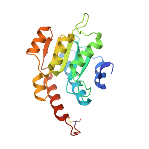
Structure guided mutagenesis reveals the substrate determinants of guanine deaminase.

Singh, J.Gaded, V.Bitra, A.Anand, R.

(2021) J Struct Biol 213: 107747-107747

  • DOI: https://doi.org/10.1016/j.jsb.2021.107747
  • Primary Citation Related Structures: 
    7C3S, 7C3T, 7C3U

  • PubMed Abstract: 

    Guanine deaminases (GDs) are essential enzymes that regulate the overall nucleobase pool. Since the deamination of guanine to xanthine results in the production of a mutagenic base, these enzymes have evolved to be very specific in nature. Surprisingly, they accept structurally distinct triazine ammeline, an intermediate in the melamine pathway, as one of the moonlighting substrates. Here, by employing NE0047 (a GD from Nitrosomonas europaea), we delineate the nuance in the catalytic mechanism that allows these two distinct substrates to be catalyzed. A combination of enzyme kinetics, X-ray crystallographic, and calorimetric studies reveal that GDs operate via a dual proton shuttle mechanism with two glutamates, E79 and E143, crucial for deamination. Additionally, N66 appears to be central for substrate anchoring and participates in catalysis. The study highlights the importance of closure of the catalytic loop and of maintenance of the hydrophobic core by capping residues like F141 and F48 for the creation of an apt environment for activation of the zinc-assisted catalysis. This study also analyzes evolutionarily distinct GDs and asserts that GDs incorporate subtle variations in the active site architectures while keeping the most critical active site determinants conserved.


  • Organizational Affiliation
    • Department of Chemistry, Indian Institute of Technology Bombay, Mumbai, Maharashtra 400076, India.

Macromolecule Content 

  • Total Structure Weight: 41.42 kDa 
  • Atom Count: 2,905 
  • Modeled Residue Count: 370 
  • Deposited Residue Count: 390 
  • Unique protein chains: 1

Macromolecules

Find similar proteins by:|  3D Structure
Entity ID: 1
MoleculeChains  Sequence LengthOrganismDetailsImage
Cytidine and deoxycytidylate deaminase zinc-binding region
A, B
195Nitrosomonas europaea ATCC 19718Mutation(s): 1 
Gene Names: NE0047
UniProt
Find proteins for Q82Y41 (Nitrosomonas europaea (strain ATCC 19718 / CIP 103999 / KCTC 2705 / NBRC 14298))
Explore Q82Y41 
Go to UniProtKB:  Q82Y41
Entity Groups
Sequence Clusters30% Identity50% Identity70% Identity90% Identity95% Identity100% Identity
UniProt GroupQ82Y41
Sequence Annotations
Expand
Reference Sequence

Small Molecules

Ligands 4 Unique
IDChains Name / Formula / InChI Key2D Diagram3D Interactions
AZG
(Subject of Investigation/LOI)

Query on AZG


Download:Ideal Coordinates CCD File
G [auth B]5-AMINO-1H-[1,2,3]TRIAZOLO[4,5-D]PYRIMIDIN-7-OL
C4 H4 N6 O
LPXQRXLUHJKZIE-UHFFFAOYSA-N
GOL

Query on GOL


Download:Ideal Coordinates CCD File
I [auth B]GLYCEROL
C3 H8 O3
PEDCQBHIVMGVHV-UHFFFAOYSA-N
ZN
(Subject of Investigation/LOI)

Query on ZN


Download:Ideal Coordinates CCD File
C [auth A],
F [auth B]
ZINC ION
Zn
PTFCDOFLOPIGGS-UHFFFAOYSA-N
EDO

Query on EDO


Download:Ideal Coordinates CCD File
D [auth A],
E [auth A],
H [auth B]
1,2-ETHANEDIOL
C2 H6 O2
LYCAIKOWRPUZTN-UHFFFAOYSA-N

Experimental Data & Validation

Experimental Data

  • Method: X-RAY DIFFRACTION
  • Resolution: 1.66 Å
  • R-Value Free:  0.193 (Depositor), 0.190 (DCC) 
  • R-Value Work:  0.160 (Depositor) 
  • R-Value Observed: 0.161 (Depositor) 
Space Group: P 21 21 21
Unit Cell:
Length ( Å )Angle ( ˚ )
a = 38.491α = 90
b = 73.629β = 90
c = 109.997γ = 90
Software Package:
Software NamePurpose
REFMACrefinement
HKL-3000data reduction
HKL-3000data scaling
MOLREPphasing

Structure Validation

View Full Validation Report


Ligand Structure Quality Assessment 


Entry History 

& Funding Information

Deposition Data


Funding OrganizationLocationGrant Number
Other governmentIndiaEMR/2015/ 002121

Revision History  (Full details and data files)

  • Version 1.0: 2021-05-26
    Type: Initial release
  • Version 1.1: 2021-06-02
    Changes: Database references
  • Version 1.2: 2021-06-16
    Changes: Database references
  • Version 1.3: 2023-11-29
    Changes: Data collection, Database references, Refinement description
  • Version 1.4: 2024-10-30
    Changes: Structure summary