7C3F | pdb_00007c3f

Crystal structure of ferredoxin: thioredoxin reductase and thioredoxin m2 complex


Experimental Data Snapshot

  • Method: X-RAY DIFFRACTION
  • Resolution: 2.40 Å
  • R-Value Free: 
    0.245 (Depositor), 0.245 (DCC) 
  • R-Value Work: 
    0.209 (Depositor), 0.210 (DCC) 
  • R-Value Observed: 
    0.211 (Depositor) 

Starting Model: experimental
View more details

wwPDB Validation 3D Report Full Report

Validation slider image for 7C3F

This is version 1.3 of the entry. See complete history

Literature

Structural basis for thioredoxin isoform-based fine-tuning of ferredoxin-thioredoxin reductase activity.

Juniar, L.Tanaka, H.Yoshida, K.Hisabori, T.Kurisu, G.

(2020) Protein Sci 29: 2538-2545

  • DOI: https://doi.org/10.1002/pro.3964
  • Primary Citation Related Structures: 
    7BZK, 7C2B, 7C3F, 7C65

  • PubMed Abstract: 

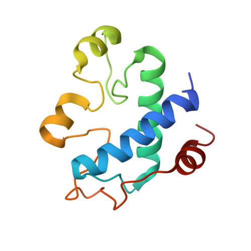
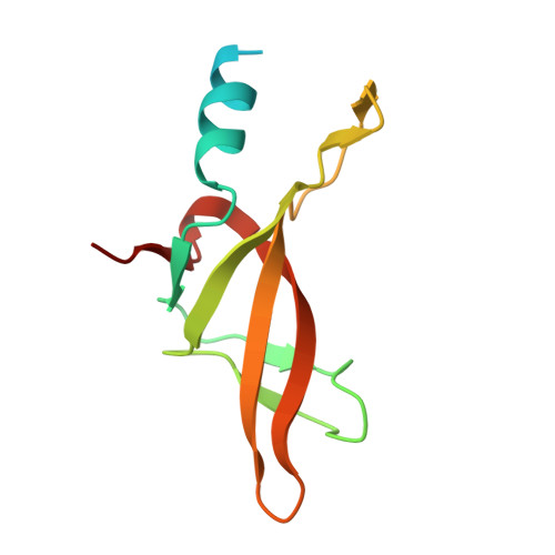
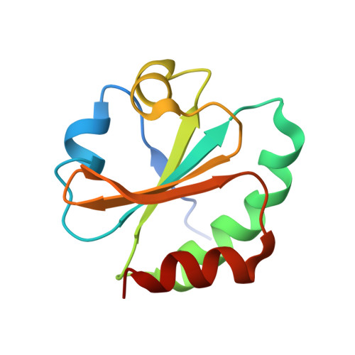
    Photosynthetic electron transport occurs on the thylakoid membrane of chloroplasts. Ferredoxin (Fd), the final acceptor in the electron transport chain, distributes electrons to several Fd-dependent enzymes including Fd-thioredoxin reductase (FTR). A cascade from Fd to FTR further reduces Thioredoxin (Trx), which tunes the activity of target metabolic enzymes eventually in a light-dependent manner. We previously reported that 10 Trx isoforms in Arabidopsis thaliana can be clustered into three classes based on the kinetics of the FTR-dependent reduction (high-, middle-, and low-efficiency classes). In this study, we determined the X-ray structure of three electron transfer complexes of FTR and Trx isoform, Trx-y1, Trx-f2, and Trx-m2, as representative examples of each class. Superposition of the FTR structure with/without Trx showed no main chain structural changes upon complex formation. There was no significant conformational change for single and complexed Trx-m structures. Nonetheless, the interface of FTR:Trx complexes displayed significant variation. Comparative analysis of the three structures showed two types of intermolecular interactions; (i) common interactions shared by all three complexes and (ii) isoform-specific interactions, which might be important for fine-tuning FTR:Trx activity. Differential electrostatic potentials of Trx isoforms may be key to isoform-specific interactions.


  • Organizational Affiliation
    • Institute for Protein Research, Osaka University, Suita, Osaka, Japan.

Macromolecule Content 

  • Total Structure Weight: 294.86 kDa 
  • Atom Count: 18,993 
  • Modeled Residue Count: 2,328 
  • Deposited Residue Count: 2,616 
  • Unique protein chains: 3

Macromolecules

Find similar proteins by:|  3D Structure
Entity ID: 1
MoleculeChains  Sequence LengthOrganismDetailsImage
Ferredoxin-thioredoxin reductase catalytic chain, chloroplastic
A,
D,
G,
J,
M,
P,
S,
W [auth V]
115Arabidopsis thalianaMutation(s): 0 
Gene Names: FTRC
EC: 1.8.7.2
UniProt
Find proteins for Q9SJ89 (Arabidopsis thaliana)
Explore Q9SJ89 
Go to UniProtKB:  Q9SJ89
Entity Groups
Sequence Clusters30% Identity50% Identity70% Identity90% Identity95% Identity100% Identity
UniProt GroupQ9SJ89
Sequence Annotations
Expand
Reference Sequence
Find similar proteins by:|  3D Structure
Entity ID: 2
MoleculeChains  Sequence LengthOrganismDetailsImage
Ferredoxin-thioredoxin reductase variable chain, chloroplastic
B, E, H, K, N
B, E, H, K, N, Q, T
112Arabidopsis thalianaMutation(s): 0 
Gene Names: FTRA2ferredoxinAt5g08410At5g08410/F8L15_140C24_LOCUS21561F8L15.14
UniProt
Find proteins for Q8LBP6 (Arabidopsis thaliana)
Explore Q8LBP6 
Go to UniProtKB:  Q8LBP6
Entity Groups
Sequence Clusters30% Identity50% Identity70% Identity90% Identity95% Identity100% Identity
UniProt GroupQ8LBP6
Sequence Annotations
Expand
Reference Sequence
Find similar proteins by:|  3D Structure
Entity ID: 3
MoleculeChains  Sequence LengthOrganismDetailsImage
Thioredoxin M2, chloroplastic
C,
F,
I,
L,
O,
R,
U,
V [auth W]
114Arabidopsis thalianaMutation(s): 1 
Gene Names: TRXM2At4g03520F9H3.15T5L23.1
UniProt
Find proteins for Q9SEU8 (Arabidopsis thaliana)
Explore Q9SEU8 
Go to UniProtKB:  Q9SEU8
Entity Groups
Sequence Clusters30% Identity50% Identity70% Identity90% Identity95% Identity100% Identity
UniProt GroupQ9SEU8
Sequence Annotations
Expand
Reference Sequence

Small Molecules

Ligands 2 Unique
IDChains Name / Formula / InChI Key2D Diagram3D Interactions
SF4
(Subject of Investigation/LOI)

Query on SF4


Download:Ideal Coordinates CCD File
AA [auth D]
DA [auth G]
FA [auth J]
IA [auth M]
LA [auth P]
AA [auth D],
DA [auth G],
FA [auth J],
IA [auth M],
LA [auth P],
NA [auth S],
X [auth A]
IRON/SULFUR CLUSTER
Fe4 S4
LJBDFODJNLIPKO-UHFFFAOYSA-N
NA

Query on NA


Download:Ideal Coordinates CCD File
BA [auth D]
CA [auth E]
EA [auth G]
GA [auth J]
HA [auth L]
BA [auth D],
CA [auth E],
EA [auth G],
GA [auth J],
HA [auth L],
JA [auth M],
KA [auth O],
MA [auth P],
Y [auth A],
Z [auth C]
SODIUM ION
Na
FKNQFGJONOIPTF-UHFFFAOYSA-N

Experimental Data & Validation

Experimental Data

  • Method: X-RAY DIFFRACTION
  • Resolution: 2.40 Å
  • R-Value Free:  0.245 (Depositor), 0.245 (DCC) 
  • R-Value Work:  0.209 (Depositor), 0.210 (DCC) 
  • R-Value Observed: 0.211 (Depositor) 
Space Group: C 1 2 1
Unit Cell:
Length ( Å )Angle ( ˚ )
a = 174.262α = 90
b = 137.209β = 90.21
c = 192.457γ = 90
Software Package:
Software NamePurpose
PHENIXrefinement
PDB_EXTRACTdata extraction
XDSdata reduction
XDSdata scaling
PHASERphasing

Structure Validation

View Full Validation Report


Entry History 

& Funding Information

Deposition Data


Funding OrganizationLocationGrant Number
Japan Society for the Promotion of Science (JSPS)Japan16H06560

Revision History  (Full details and data files)

  • Version 1.0: 2020-10-14
    Type: Initial release
  • Version 1.1: 2020-12-02
    Changes: Database references
  • Version 1.2: 2023-11-29
    Changes: Data collection, Database references, Refinement description
  • Version 1.3: 2024-11-06
    Changes: Structure summary