7BZ5 | pdb_00007bz5

Structure of COVID-19 virus spike receptor-binding domain complexed with a neutralizing antibody


Experimental Data Snapshot

  • Method: X-RAY DIFFRACTION
  • Resolution: 1.84 Å
  • R-Value Free: 
    0.191 (Depositor), 0.174 (DCC) 
  • R-Value Work: 
    0.167 (Depositor), 0.170 (DCC) 
  • R-Value Observed: 
    0.168 (Depositor) 

Starting Models: experimental
View more details

wwPDB Validation 3D Report Full Report

Validation slider image for 7BZ5

Ligand Structure Quality Assessment 


This is version 1.6 of the entry. See complete history

Literature

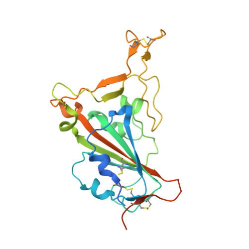
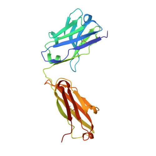
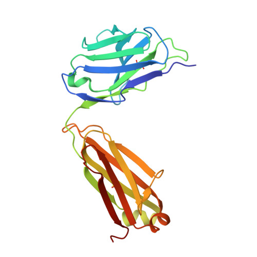
A noncompeting pair of human neutralizing antibodies block COVID-19 virus binding to its receptor ACE2.

Wu, Y.Wang, F.Shen, C.Peng, W.Li, D.Zhao, C.Li, Z.Li, S.Bi, Y.Yang, Y.Gong, Y.Xiao, H.Fan, Z.Tan, S.Wu, G.Tan, W.Lu, X.Fan, C.Wang, Q.Liu, Y.Zhang, C.Qi, J.Gao, G.F.Gao, F.Liu, L.

(2020) Science 368: 1274-1278

  • DOI: https://doi.org/10.1126/science.abc2241
  • Primary Citation Related Structures: 
    7BZ5

  • PubMed Abstract: 

    Neutralizing antibodies could potentially be used as antivirals against the coronavirus disease 2019 (COVID-19) pandemic. Here, we report isolation of four human-origin monoclonal antibodies from a convalescent patient, all of which display neutralization abilities. The antibodies B38 and H4 block binding between the spike glycoprotein receptor binding domain (RBD) of the virus and the cellular receptor angiotensin-converting enzyme 2 (ACE2). A competition assay indicated different epitopes on the RBD for these two antibodies, making them a potentially promising virus-targeting monoclonal antibody pair for avoiding immune escape in future clinical applications. Moreover, a therapeutic study in a mouse model validated that these antibodies can reduce virus titers in infected lungs. The RBD-B38 complex structure revealed that most residues on the epitope overlap with the RBD-ACE2 binding interface, explaining the blocking effect and neutralizing capacity. Our results highlight the promise of antibody-based therapeutics and provide a structural basis for rational vaccine design.


  • Organizational Affiliation
    • Department of Pathogen Microbiology, School of Basic Medical Sciences, Capital Medical University, Beijing, China. wuy@biols.ac.cn gaofeng@tib.cas.cn gaof@im.ac.cn liulei3322@aliyun.com.

Macromolecule Content 

  • Total Structure Weight: 73.36 kDa 
  • Atom Count: 5,367 
  • Modeled Residue Count: 628 
  • Deposited Residue Count: 669 
  • Unique protein chains: 3

Macromolecules

Find similar proteins by:|  3D Structure
Entity ID: 1
MoleculeChains  Sequence LengthOrganismDetailsImage
Spike protein S1229Severe acute respiratory syndrome coronavirus 2Mutation(s): 0 
Gene Names: S2
UniProt
Find proteins for P0DTC2 (Severe acute respiratory syndrome coronavirus 2)
Explore P0DTC2 
Go to UniProtKB:  P0DTC2
Entity Groups
Sequence Clusters30% Identity50% Identity70% Identity90% Identity95% Identity100% Identity
UniProt GroupP0DTC2
Glycosylation
Glycosylation Sites: 1Go to GlyGen: P0DTC2-1
Sequence Annotations
Expand
Reference Sequence
Find similar proteins by:|  3D Structure
Entity ID: 2
MoleculeChains  Sequence LengthOrganismDetailsImage
Heavy chain of B38B [auth H]222Homo sapiensMutation(s): 0 
Find similar proteins by:|  3D Structure
Entity ID: 3
MoleculeChains  Sequence LengthOrganismDetailsImage
Light chain of B38C [auth L]218Homo sapiensMutation(s): 0 

Small Molecules

Ligands 1 Unique
IDChains Name / Formula / InChI Key2D Diagram3D Interactions
NAG

Query on NAG


Download:Ideal Coordinates CCD File
D [auth A]2-acetamido-2-deoxy-beta-D-glucopyranose
C8 H15 N O6
OVRNDRQMDRJTHS-FMDGEEDCSA-N

Experimental Data & Validation

Experimental Data

  • Method: X-RAY DIFFRACTION
  • Resolution: 1.84 Å
  • R-Value Free:  0.191 (Depositor), 0.174 (DCC) 
  • R-Value Work:  0.167 (Depositor), 0.170 (DCC) 
  • R-Value Observed: 0.168 (Depositor) 
Space Group: C 1 2 1
Unit Cell:
Length ( Å )Angle ( ˚ )
a = 195.226α = 90
b = 86.417β = 100.19
c = 56.827γ = 90
Software Package:
Software NamePurpose
HKL-2000data scaling
PHENIXrefinement
PDB_EXTRACTdata extraction
HKL-2000data reduction
PHASERphasing

Structure Validation

View Full Validation Report


Ligand Structure Quality Assessment 


Entry History 

& Funding Information

Deposition Data


Funding OrganizationLocationGrant Number
National Natural Science Foundation of China (NSFC)China31872745, 81902058

Revision History  (Full details and data files)

  • Version 1.0: 2020-05-13
    Type: Initial release
  • Version 1.1: 2020-05-27
    Changes: Database references
  • Version 1.2: 2020-06-24
    Changes: Database references
  • Version 1.3: 2020-07-29
    Type: Remediation
    Reason: Carbohydrate remediation
    Changes: Data collection, Derived calculations, Structure summary
  • Version 1.4: 2021-03-10
    Changes: Structure summary
  • Version 1.5: 2023-11-29
    Changes: Data collection, Database references, Refinement description
  • Version 1.6: 2024-10-30
    Changes: Structure summary