7BYU | pdb_00007byu

Crystal structure of Acidovorax avenae L-fucose mutarotase (apo form)


Experimental Data Snapshot

  • Method: X-RAY DIFFRACTION
  • Resolution: 2.21 Å
  • R-Value Free: 
    0.240 (Depositor), 0.242 (DCC) 
  • R-Value Work: 
    0.217 (Depositor), 0.223 (DCC) 
  • R-Value Observed: 
    0.218 (Depositor) 

Starting Model: experimental
View more details

wwPDB Validation 3D Report Full Report

Validation slider image for 7BYU

This is version 1.3 of the entry. See complete history

Literature

Functional and structural characterization of a novel L-fucose mutarotase involved in non-phosphorylative pathway of L-fucose metabolism.

Watanabe, Y.Watanabe, S.Fukui, Y.Nishiwaki, H.

(2020) Biochem Biophys Res Commun 528: 21-27

  • DOI: https://doi.org/10.1016/j.bbrc.2020.05.094
  • Primary Citation Related Structures: 
    7BYU, 7BYW

  • PubMed Abstract: 

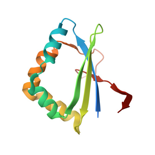
    Mutarotases catalyze the α-β anomeric conversion of monosaccharide, and play a key role in utilizing sugar as enzymes involved in sugar metabolism have specificity for the α- or β-anomer. In spite of the sequential similarity to l-rhamnose mutarotase protein superfamily (COG3254: RhaM), the ACAV_RS08160 gene in Acidovorax avenae ATCC 19860 (AaFucM) is located in a gene cluster related to non-phosphorylative l-fucose and l-galactose metabolism, and transcriptionally induced by these carbon sources; therefore, the physiological role remains unclear. Here, we report that AaFucM possesses mutarotation activity only toward l-fucose by saturation difference (SD) NMR experiments. Moreover, we determined the crystal structures of AaFucM in the apo form and in the l-fucose-bound form at resolutions of 2.21 and 1.75 Å, respectively. The overall structural folding was clearly similar to the RhaM members, differed from the known l-fucose mutarotase (COG4154: FucU), strongly indicating their convergent evolution. The structure-based mutational analyses suggest that Tyr18 is important for catalytic action, and that Gln87 and Trp99 are involved in the l-fucose-specific recognition.


  • Organizational Affiliation
    • Department of Bioscience, Graduate School of Agriculture, Ehime University, 3-5-7 Tarumi, Matsuyama, Ehime, 790-8566, Japan; Faculty of Agriculture, Ehime University, 3-5-7 Tarumi, Matsuyama, Ehime, 790-8566, Japan.

Macromolecule Content 

  • Total Structure Weight: 28.92 kDa 
  • Atom Count: 1,918 
  • Modeled Residue Count: 216 
  • Deposited Residue Count: 240 
  • Unique protein chains: 1

Macromolecules

Find similar proteins by:|  3D Structure
Entity ID: 1
MoleculeChains  Sequence LengthOrganismDetailsImage
L-fucose mutarotase
A, B
120Paracidovorax avenae ATCC 19860Mutation(s): 0 
Gene Names: Acav_1655
UniProt
Find proteins for F0Q4R9 (Paracidovorax avenae (strain ATCC 19860 / DSM 7227 / CCUG 15838 / JCM 20985 / LMG 2117 / NCPPB 1011))
Explore F0Q4R9 
Go to UniProtKB:  F0Q4R9
Entity Groups
Sequence Clusters30% Identity50% Identity70% Identity90% Identity95% Identity100% Identity
UniProt GroupF0Q4R9
Sequence Annotations
Expand
Reference Sequence

Experimental Data & Validation

Experimental Data

  • Method: X-RAY DIFFRACTION
  • Resolution: 2.21 Å
  • R-Value Free:  0.240 (Depositor), 0.242 (DCC) 
  • R-Value Work:  0.217 (Depositor), 0.223 (DCC) 
  • R-Value Observed: 0.218 (Depositor) 
Space Group: P 31
Unit Cell:
Length ( Å )Angle ( ˚ )
a = 69.465α = 90
b = 69.465β = 90
c = 69.2γ = 120
Software Package:
Software NamePurpose
XDSdata processing
PHENIXrefinement
PDB_EXTRACTdata extraction
Cootmodel building
PHASERphasing
Aimlessdata scaling

Structure Validation

View Full Validation Report


Entry History 

Deposition Data

Revision History  (Full details and data files)

  • Version 1.0: 2020-05-27
    Type: Initial release
  • Version 1.1: 2020-06-10
    Changes: Database references
  • Version 1.2: 2020-06-24
    Changes: Database references
  • Version 1.3: 2023-11-29
    Changes: Data collection, Database references, Refinement description