7BYT | pdb_00007byt

Crystal structure of exo-beta-1,3-galactanase from Phanerochaete chrysosporium Pc1,3Gal43A with galactose


Experimental Data Snapshot

  • Method: X-RAY DIFFRACTION
  • Resolution: 1.50 Å
  • R-Value Free: 
    0.160 (Depositor), 0.159 (DCC) 
  • R-Value Work: 
    0.134 (Depositor), 0.134 (DCC) 

Starting Model: experimental
View more details

wwPDB Validation 3D Report Full Report

Validation slider image for 7BYT

Ligand Structure Quality Assessment 


This is version 1.3 of the entry. See complete history

Literature

Unique active-site and subsite features in the arabinogalactan-degrading GH43 exo-beta-1,3-galactanase from Phanerochaete chrysosporium .

Matsuyama, K.Kishine, N.Fujimoto, Z.Sunagawa, N.Kotake, T.Tsumuraya, Y.Samejima, M.Igarashi, K.Kaneko, S.

(2020) J Biological Chem 295: 18539-18552

  • DOI: https://doi.org/10.1074/jbc.RA120.016149
  • Primary Citation Related Structures: 
    7BYS, 7BYT, 7BYV, 7BYX

  • PubMed Abstract: 

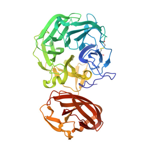
    Arabinogalactan proteins (AGPs) are plant proteoglycans with functions in growth and development. However, these functions are largely unexplored, mainly because of the complexity of the sugar moieties. These carbohydrate sequences are generally analyzed with the aid of glycoside hydrolases. The exo-β-1,3-galactanase is a glycoside hydrolase from the basidiomycete Phanerochaete chrysosporium ( Pc 1,3Gal43A), which specifically cleaves AGPs. However, its structure is not known in relation to its mechanism bypassing side chains. In this study, we solved the apo and liganded structures of Pc 1,3Gal43A, which reveal a glycoside hydrolase family 43 subfamily 24 (GH43_sub24) catalytic domain together with a carbohydrate-binding module family 35 (CBM35) binding domain. GH43_sub24 is known to lack the catalytic base Asp conserved among other GH43 subfamilies. Our structure in combination with kinetic analyses reveals that the tautomerized imidic acid group of Gln 263 serves as the catalytic base residue instead. Pc 1,3Gal43A has three subsites that continue from the bottom of the catalytic pocket to the solvent. Subsite -1 contains a space that can accommodate the C-6 methylol of Gal, enabling the enzyme to bypass the β-1,6-linked galactan side chains of AGPs. Furthermore, the galactan-binding domain in CBM35 has a different ligand interaction mechanism from other sugar-binding CBM35s, including those that bind galactomannan. Specifically, we noted a Gly → Trp substitution, which affects pyranose stacking, and an Asp → Asn substitution in the binding pocket, which recognizes β-linked rather than α-linked Gal residues. These findings should facilitate further structural analysis of AGPs and may also be helpful in engineering designer enzymes for efficient biomass utilization.


  • Organizational Affiliation
    • Department of Biomaterial Sciences, Graduate School of Agricultural and Life Sciences, University of Tokyo, Tokyo, Japan.

Macromolecule Content 

  • Total Structure Weight: 47.41 kDa 
  • Atom Count: 3,923 
  • Modeled Residue Count: 427 
  • Deposited Residue Count: 427 
  • Unique protein chains: 1

Macromolecules

Find similar proteins by:|  3D Structure
Entity ID: 1
MoleculeChains  Sequence LengthOrganismDetailsImage
Galactan 1,3-beta-galactosidase427Phanerodontia chrysosporiumMutation(s): 0 
EC: 3.2.1.145
UniProt
Find proteins for Q50KB2 (Phanerodontia chrysosporium)
Explore Q50KB2 
Go to UniProtKB:  Q50KB2
Entity Groups
Sequence Clusters30% Identity50% Identity70% Identity90% Identity95% Identity100% Identity
UniProt GroupQ50KB2
Glycosylation
Glycosylation Sites: 3
Sequence Annotations
Expand
Reference Sequence

Oligosaccharides

Help  
Entity ID: 2
MoleculeChains Length2D Diagram GlycosylationD Interactions
2-acetamido-2-deoxy-beta-D-glucopyranose-(1-4)-2-acetamido-2-deoxy-beta-D-glucopyranose
B
2N-Glycosylation
Glycosylation Resources
GlyTouCan: G42666HT
GlyCosmos: G42666HT
GlyGen: G42666HT

Small Molecules

Ligands 7 Unique
IDChains Name / Formula / InChI Key2D Diagram3D Interactions
NAG

Query on NAG


Download:Ideal Coordinates CCD File
C [auth A],
D [auth A]
2-acetamido-2-deoxy-beta-D-glucopyranose
C8 H15 N O6
OVRNDRQMDRJTHS-FMDGEEDCSA-N
GAL
(Subject of Investigation/LOI)

Query on GAL


Download:Ideal Coordinates CCD File
G [auth A]beta-D-galactopyranose
C6 H12 O6
WQZGKKKJIJFFOK-FPRJBGLDSA-N
GLA
(Subject of Investigation/LOI)

Query on GLA


Download:Ideal Coordinates CCD File
F [auth A]alpha-D-galactopyranose
C6 H12 O6
WQZGKKKJIJFFOK-PHYPRBDBSA-N
GOL

Query on GOL


Download:Ideal Coordinates CCD File
L [auth A],
M [auth A],
N [auth A],
O [auth A]
GLYCEROL
C3 H8 O3
PEDCQBHIVMGVHV-UHFFFAOYSA-N
ACY

Query on ACY


Download:Ideal Coordinates CCD File
H [auth A],
I [auth A]
ACETIC ACID
C2 H4 O2
QTBSBXVTEAMEQO-UHFFFAOYSA-N
ACT

Query on ACT


Download:Ideal Coordinates CCD File
J [auth A],
K [auth A]
ACETATE ION
C2 H3 O2
QTBSBXVTEAMEQO-UHFFFAOYSA-M
CA

Query on CA


Download:Ideal Coordinates CCD File
E [auth A]CALCIUM ION
Ca
BHPQYMZQTOCNFJ-UHFFFAOYSA-N

Experimental Data & Validation

Experimental Data

  • Method: X-RAY DIFFRACTION
  • Resolution: 1.50 Å
  • R-Value Free:  0.160 (Depositor), 0.159 (DCC) 
  • R-Value Work:  0.134 (Depositor), 0.134 (DCC) 
Space Group: P 21 21 21
Unit Cell:
Length ( Å )Angle ( ˚ )
a = 50.818α = 90
b = 66.565β = 90
c = 106.39γ = 90
Software Package:
Software NamePurpose
PHENIXrefinement
REFMACrefinement
Cootmodel building
DENZOdata reduction
SCALEPACKdata scaling
MOLREPphasing

Structure Validation

View Full Validation Report


Ligand Structure Quality Assessment 


Entry History 

& Funding Information

Deposition Data


Funding OrganizationLocationGrant Number
Japan Society for the Promotion of Science (JSPS)Japan18H05494

Revision History  (Full details and data files)

  • Version 1.0: 2020-11-04
    Type: Initial release
  • Version 1.1: 2021-01-13
    Changes: Database references
  • Version 1.2: 2024-04-03
    Changes: Data collection, Database references, Refinement description
  • Version 1.3: 2024-10-30
    Changes: Structure summary