


Structure of subunit I of the anthranilate synthase complex of Mycolicibacterium smegmatis
Chen, Y., Jia, H., Liu, R., Che, S., Zhang, Q., Bartlam, M.(2020) Biochem Biophys Res Commun 527: 37-41
Experimental Data Snapshot
Starting Model: experimental
View more details
Entity ID: 1 | |||||
|---|---|---|---|---|---|
| Molecule | Chains | Sequence Length | Organism | Details | Image |
| Anthranilate synthase component 1 | 524 | Mycolicibacterium smegmatis | Mutation(s): 0 Gene Names: trpE, ERS451418_03206 EC: 4.1.3.27 | 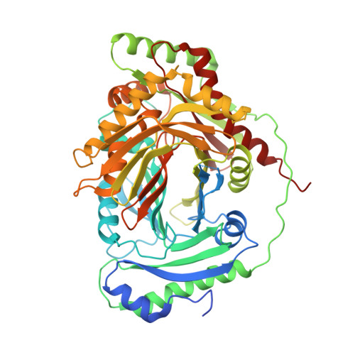 | |
UniProt | |||||
Entity Groups | |||||
| Sequence Clusters | 30% Identity50% Identity70% Identity90% Identity95% Identity100% Identity | ||||
| UniProt Group | A0QX93 | ||||
Sequence AnnotationsExpand | |||||
Reference Sequence | |||||
| Ligands 4 Unique | |||||
|---|---|---|---|---|---|
| ID | Chains | Name / Formula / InChI Key | 2D Diagram | 3D Interactions | |
| BEZ (Subject of Investigation/LOI) Download:Ideal Coordinates CCD File | C [auth A], I [auth B] | BENZOIC ACID C7 H6 O2 WPYMKLBDIGXBTP-UHFFFAOYSA-N |  | ||
| GOL Download:Ideal Coordinates CCD File | G [auth A], H [auth A], K [auth B], L [auth B] | GLYCEROL C3 H8 O3 PEDCQBHIVMGVHV-UHFFFAOYSA-N |  | ||
| PYR Download:Ideal Coordinates CCD File | F [auth A] | PYRUVIC ACID C3 H4 O3 LCTONWCANYUPML-UHFFFAOYSA-N |  | ||
| MG Download:Ideal Coordinates CCD File | D [auth A], E [auth A], J [auth B] | MAGNESIUM ION Mg JLVVSXFLKOJNIY-UHFFFAOYSA-N |  | ||
| Length ( Å ) | Angle ( ˚ ) |
|---|---|
| a = 60.822 | α = 81.88 |
| b = 66.076 | β = 72.73 |
| c = 80.718 | γ = 70.88 |
| Software Name | Purpose |
|---|---|
| PHENIX | refinement |
| HKL-3000 | data reduction |
| HKL-3000 | data scaling |
| PHENIX | phasing |
| Funding Organization | Location | Grant Number |
|---|---|---|
| National Natural Science Foundation of China (NSFC) | China | 31800627 |


