7BUQ | pdb_00007buq

mcGAS bound with 23-cGAMP


Experimental Data Snapshot

  • Method: X-RAY DIFFRACTION
  • Resolution: 3.09 Å
  • R-Value Free: 
    0.296 (Depositor), 0.293 (DCC) 
  • R-Value Work: 
    0.249 (Depositor), 0.249 (DCC) 
  • R-Value Observed: 
    0.254 (Depositor) 

Starting Model: experimental
View more details

wwPDB Validation 3D Report Full Report

Validation slider image for 7BUQ

Ligand Structure Quality Assessment 


This is version 1.1 of the entry. See complete history

Literature

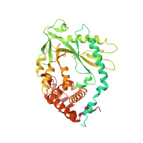
Mn2+Directly Activates cGAS and Structural Analysis Suggests Mn2+Induces a Noncanonical Catalytic Synthesis of 2'3'-cGAMP.

Zhao, Z.Ma, Z.Wang, B.Guan, Y.Su, X.D.Jiang, Z.

(2020) Cell Rep 32: 108053-108053

  • DOI: https://doi.org/10.1016/j.celrep.2020.108053
  • Primary Citation Related Structures: 
    7BUJ, 7BUM, 7BUQ

  • PubMed Abstract: 

    DNA binding allosterically activates the cytosolic DNA sensor cGAS (cyclic GMP-AMP [cGAMP] synthase) to synthesize 2'3'-cGAMP, using Mg 2+ as the metal cofactor that catalyzes two nucleotidyl-transferring reactions. We previously found that Mn 2+ potentiates cGAS activation, but the underlying mechanism remains unclear. Here, we report that Mn 2+ directly activates cGAS. Structural analysis reveals that Mn 2+ -activated cGAS undergoes globally similar conformational changes to DNA-activated cGAS but forms a unique η1 helix to widen the catalytic pocket, allowing substrate entry and cGAMP synthesis. Strikingly, in Mn 2+ -activated cGAS, the linear intermediates pppGpG and pGpA take an inverted orientation in the active pocket, suggesting a noncanonical but accelerated cGAMP cyclization without substrate flip-over. Moreover, unlike the octahedral coordination around Mg 2+ , the two catalytic Mn 2+ are coordinated by triphosphate moiety of the inverted substrate, independent of the catalytic triad residues. Our findings thus uncover Mn 2+ as a cGAS activator that initiates noncanonical 2'3'-cGAMP synthesis.


  • Organizational Affiliation
    • Key Laboratory of Cell Proliferation and Differentiation of the Ministry of Education, School of Life Sciences, Peking University, Beijing 100871, China; Peking-Tsinghua Center for Life Sciences, Peking University, Beijing 100871, China.

Macromolecule Content 

  • Total Structure Weight: 118.08 kDa 
  • Atom Count: 6,070 
  • Modeled Residue Count: 723 
  • Deposited Residue Count: 1,014 
  • Unique protein chains: 1

Macromolecules

Find similar proteins by:|  3D Structure
Entity ID: 1
MoleculeChains  Sequence LengthOrganismDetailsImage
Cyclic GMP-AMP synthase
A, B
507Mus musculusMutation(s): 0 
Gene Names: CgasMb21d1
EC: 2.7.7.86
UniProt & NIH Common Fund Data Resources
Find proteins for Q8C6L5 (Mus musculus)
Explore Q8C6L5 
Go to UniProtKB:  Q8C6L5
IMPC:  MGI:2442261
Entity Groups
Sequence Clusters30% Identity50% Identity70% Identity90% Identity95% Identity100% Identity
UniProt GroupQ8C6L5
Sequence Annotations
Expand
Reference Sequence

Experimental Data & Validation

Experimental Data

  • Method: X-RAY DIFFRACTION
  • Resolution: 3.09 Å
  • R-Value Free:  0.296 (Depositor), 0.293 (DCC) 
  • R-Value Work:  0.249 (Depositor), 0.249 (DCC) 
  • R-Value Observed: 0.254 (Depositor) 
Space Group: P 1 21 1
Unit Cell:
Length ( Å )Angle ( ˚ )
a = 47.645α = 90
b = 109.812β = 94.13
c = 75.637γ = 90
Software Package:
Software NamePurpose
PHENIXrefinement
HKL-2000data reduction
SCALEPACKdata scaling
PHASERphasing
PDB_EXTRACTdata extraction

Structure Validation

View Full Validation Report


Ligand Structure Quality Assessment 


Entry History 

& Funding Information

Deposition Data


Funding OrganizationLocationGrant Number
National Science Foundation (NSF, China)China31670740
National Science Foundation (NSF, China)China31270803

Revision History  (Full details and data files)

  • Version 1.0: 2020-09-02
    Type: Initial release
  • Version 1.1: 2023-11-29
    Changes: Data collection, Database references, Refinement description