7BIU | pdb_00007biu

XFEL crystal structure of cytochrome c peroxidase compound II


Experimental Data Snapshot

  • Method: X-RAY DIFFRACTION
  • Resolution: 1.06 Å
  • R-Value Work: 
    0.140 (Depositor), 0.148 (DCC) 

Starting Model: experimental
View more details

wwPDB Validation 3D Report Full Report

Validation slider image for 7BIU

Ligand Structure Quality Assessment 


This is version 1.2 of the entry. See complete history

Literature

XFEL Crystal Structures of Peroxidase Compound II.

Kwon, H.Basran, J.Pathak, C.Hussain, M.Freeman, S.L.Fielding, A.J.Bailey, A.J.Stefanou, N.Sparkes, H.A.Tosha, T.Yamashita, K.Hirata, K.Murakami, H.Ueno, G.Ago, H.Tono, K.Yamamoto, M.Sawai, H.Shiro, Y.Sugimoto, H.Raven, E.L.Moody, P.C.E.

(2021) Angew Chem Int Ed Engl 60: 14578-14585

  • DOI: https://doi.org/10.1002/anie.202103010
  • Primary Citation Related Structures: 
    7BI1, 7BIU

  • PubMed Abstract: 

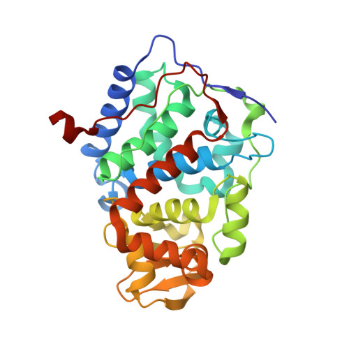
    Oxygen activation in all heme enzymes requires the formation of high oxidation states of iron, usually referred to as ferryl heme. There are two known intermediates: Compound I and Compound II. The nature of the ferryl heme-and whether it is an Fe IV =O or Fe IV -OH species-is important for controlling reactivity across groups of heme enzymes. The most recent evidence for Compound I indicates that the ferryl heme is an unprotonated Fe IV =O species. For Compound II, the nature of the ferryl heme is not unambiguously established. Here, we report 1.06 Å and 1.50 Å crystal structures for Compound II intermediates in cytochrome c peroxidase (CcP) and ascorbate peroxidase (APX), collected using the X-ray free electron laser at SACLA. The structures reveal differences between the two peroxidases. The iron-oxygen bond length in CcP (1.76 Å) is notably shorter than in APX (1.87 Å). The results indicate that the ferryl species is finely tuned across Compound I and Compound II species in closely related peroxidase enzymes. We propose that this fine-tuning is linked to the functional need for proton delivery to the heme.


  • Organizational Affiliation
    • School of Chemistry, University of Bristol, Cantock's Close, Bristol, BS8 1TS, UK.

Macromolecule Content 

  • Total Structure Weight: 34.25 kDa 
  • Atom Count: 3,104 
  • Modeled Residue Count: 292 
  • Deposited Residue Count: 294 
  • Unique protein chains: 1

Macromolecules

Find similar proteins by:|  3D Structure
Entity ID: 1
MoleculeChains  Sequence LengthOrganismDetailsImage
Cytochrome c peroxidase, mitochondrial294Saccharomyces cerevisiaeMutation(s): 0 
Gene Names: SCNYR20_0010026000SCP684_0010026400
EC: 1.11.1.5
UniProt
Find proteins for P00431 (Saccharomyces cerevisiae (strain ATCC 204508 / S288c))
Explore P00431 
Go to UniProtKB:  P00431
Entity Groups
Sequence Clusters30% Identity50% Identity70% Identity90% Identity95% Identity100% Identity
UniProt GroupP00431
Sequence Annotations
Expand
Reference Sequence

Small Molecules

Ligands 1 Unique
IDChains Name / Formula / InChI Key2D Diagram3D Interactions
HEC

Query on HEC


Download:Ideal Coordinates CCD File
B [auth A]HEME C
C34 H34 Fe N4 O4
HXQIYSLZKNYNMH-LJNAALQVSA-N

Experimental Data & Validation

Experimental Data

  • Method: X-RAY DIFFRACTION
  • Resolution: 1.06 Å
  • R-Value Work:  0.140 (Depositor), 0.148 (DCC) 
Space Group: P 21 21 21
Unit Cell:
Length ( Å )Angle ( ˚ )
a = 50.81α = 90
b = 75.54β = 90
c = 106.57γ = 90
Software Package:
Software NamePurpose
SHELXrefinement
PDB_EXTRACTdata extraction
CrystFELdata reduction
CrystFELdata scaling
PHENIXphasing

Structure Validation

View Full Validation Report


Ligand Structure Quality Assessment 


Entry History 

& Funding Information

Deposition Data


Funding OrganizationLocationGrant Number
Biotechnology and Biological Sciences Research Council (BBSRC)United KingdomBB/N015940/1

Revision History  (Full details and data files)

  • Version 1.0: 2021-04-21
    Type: Initial release
  • Version 1.1: 2021-06-30
    Changes: Database references
  • Version 1.2: 2024-01-31
    Changes: Data collection, Database references, Refinement description