7BH9

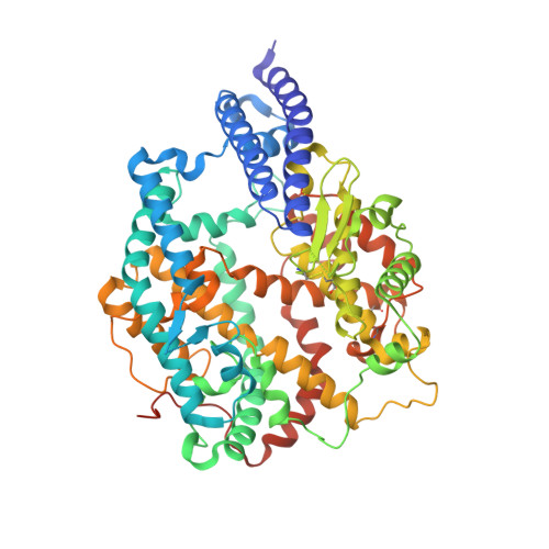
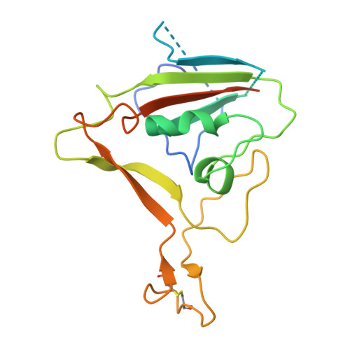
SARS-CoV-2 RBD-62 in complex with ACE2 peptidase domain


Experimental Data Snapshot

  • Method: ELECTRON MICROSCOPY
  • Resolution: 2.90 Å
  • Aggregation State: PARTICLE 
  • Reconstruction Method: SINGLE PARTICLE 

Starting Model: experimental
View more details

wwPDB Validation   3D Report Full Report


This is version 1.4 of the entry. See complete history


Literature

SARS-CoV-2 variant prediction and antiviral drug design are enabled by RBD in vitro evolution.

Zahradnik, J.Marciano, S.Shemesh, M.Zoler, E.Harari, D.Chiaravalli, J.Meyer, B.Rudich, Y.Li, C.Marton, I.Dym, O.Elad, N.Lewis, M.G.Andersen, H.Gagne, M.Seder, R.A.Douek, D.C.Schreiber, G.

(2021) Nat Microbiol 6: 1188-1198

  • DOI: https://doi.org/10.1038/s41564-021-00954-4
  • Primary Citation of Related Structures:  
    7BH9

  • PubMed Abstract: 

    SARS-CoV-2 variants of interest and concern will continue to emerge for the duration of the COVID-19 pandemic. To map mutations in the receptor-binding domain (RBD) of the spike protein that affect binding to angiotensin-converting enzyme 2 (ACE2), the receptor for SARS-CoV-2, we applied in vitro evolution to affinity-mature the RBD. Multiple rounds of random mutagenic libraries of the RBD were sorted against decreasing concentrations of ACE2, resulting in the selection of higher affinity RBD binders. We found that mutations present in more transmissible viruses (S477N, E484K and N501Y) were preferentially selected in our high-throughput screen. Evolved RBD mutants include prominently the amino acid substitutions found in the RBDs of B.1.620, B.1.1.7 (Alpha), B1.351 (Beta) and P.1 (Gamma) variants. Moreover, the incidence of RBD mutations in the population as presented in the GISAID database (April 2021) is positively correlated with increased binding affinity to ACE2. Further in vitro evolution increased binding by 1,000-fold and identified mutations that may be more infectious if they evolve in the circulating viral population, for example, Q498R is epistatic to N501Y. We show that our high-affinity variant RBD-62 can be used as a drug to inhibit infection with SARS-CoV-2 and variants Alpha, Beta and Gamma in vitro. In a model of SARS-CoV-2 challenge in hamster, RBD-62 significantly reduced clinical disease when administered before or after infection. A 2.9 Å cryo-electron microscopy structure of the high-affinity complex of RBD-62 and ACE2, including all rapidly spreading mutations, provides a structural basis for future drug and vaccine development and for in silico evaluation of known antibodies.


  • Organizational Affiliation

    Department of Biomolecular Sciences, Weizmann Institute of Science, Rehovot, Israel.


Macromolecules
Find similar proteins by:  (by identity cutoff)  |  3D Structure
Entity ID: 1
MoleculeChains Sequence LengthOrganismDetailsImage
Angiotensin-converting enzyme 2610Homo sapiensMutation(s): 0 
Gene Names: ACE2UNQ868/PRO1885
EC: 3.4.17.23 (PDB Primary Data), 3.4.17 (PDB Primary Data)
UniProt & NIH Common Fund Data Resources
Find proteins for Q9BYF1 (Homo sapiens)
Explore Q9BYF1 
Go to UniProtKB:  Q9BYF1
PHAROS:  Q9BYF1
GTEx:  ENSG00000130234 
Entity Groups  
Sequence Clusters30% Identity50% Identity70% Identity90% Identity95% Identity100% Identity
UniProt GroupQ9BYF1
Glycosylation
Glycosylation Sites: 3Go to GlyGen: Q9BYF1-1
Sequence Annotations
Expand
  • Reference Sequence
Find similar proteins by:  (by identity cutoff)  |  3D Structure
Entity ID: 2
MoleculeChains Sequence LengthOrganismDetailsImage
Spike protein S1B [auth E]202Severe acute respiratory syndrome coronavirus 2Mutation(s): 9 
Gene Names: S2
UniProt
Find proteins for P0DTC2 (Severe acute respiratory syndrome coronavirus 2)
Explore P0DTC2 
Go to UniProtKB:  P0DTC2
Entity Groups  
Sequence Clusters30% Identity50% Identity70% Identity90% Identity95% Identity100% Identity
UniProt GroupP0DTC2
Sequence Annotations
Expand
  • Reference Sequence
Experimental Data & Validation

Experimental Data

  • Method: ELECTRON MICROSCOPY
  • Resolution: 2.90 Å
  • Aggregation State: PARTICLE 
  • Reconstruction Method: SINGLE PARTICLE 
EM Software:
TaskSoftware PackageVersion
RECONSTRUCTIONcryoSPARC3.0.1
MODEL REFINEMENTPHENIX

Structure Validation

View Full Validation Report


Entry History & Funding Information

Deposition Data


Funding OrganizationLocationGrant Number
Israel Science FoundationIsrael3814/19
Israel Science FoundationIsrael1268/18

Revision History  (Full details and data files)

  • Version 1.0: 2021-02-17
    Type: Initial release
  • Version 1.1: 2021-03-10
    Changes: Database references, Source and taxonomy, Structure summary
  • Version 1.2: 2021-09-01
    Changes: Database references
  • Version 1.3: 2021-09-22
    Changes: Data collection, Database references
  • Version 1.4: 2024-10-09
    Changes: Data collection, Refinement description, Structure summary