7BGT | pdb_00007bgt

Mason-Pfizer Monkey Virus Protease mutant C7A/D26N/C106A in complex with peptidomimetic inhibitor


Experimental Data Snapshot

  • Method: X-RAY DIFFRACTION
  • Resolution: 1.93 Å
  • R-Value Free: 
    0.212 (Depositor), 0.213 (DCC) 
  • R-Value Work: 
    0.177 (Depositor), 0.179 (DCC) 
  • R-Value Observed: 
    0.178 (Depositor) 

Starting Model: experimental
View more details

wwPDB Validation 3D Report Full Report

Validation slider image for 7BGT

This is version 1.2 of the entry. See complete history

Literature

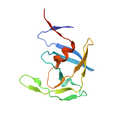
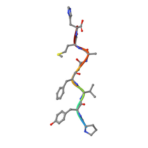
Crystal structures of inhibitor complexes of M-PMV protease with visible flap loops.

Wosicki, S.Kazmierczyk, M.Gilski, M.Zabranska, H.Pichova, I.Jaskolski, M.

(2021) Protein Sci 30: 1258-1263

  • DOI: https://doi.org/10.1002/pro.4072
  • Primary Citation Related Structures: 
    7BGT, 7BGU

  • PubMed Abstract: 

    Mason-Pfizer monkey virus protease (PR) was crystallized in complex with two pepstatin-based inhibitors in P1 space group. In both crystal structures, the extended flap loops that lock the inhibitor/substrate over the active site, are visible in the electron density either completely or with only small gaps, providing the first observation of the conformation of the flap loops in dimeric complex form of this retropepsin. The H-bond network in the active site (with D26N mutation) differs from that reported for the P2 1 crystal structures and is similar to a rarely occurring system in HIV-1 PR.


  • Organizational Affiliation
    • Center for Biocrystallographic Research, Institute of Bioorganic Chemistry, Polish Academy of Sciences, Poznan, Poland.

Macromolecule Content 

  • Total Structure Weight: 54.07 kDa 
  • Atom Count: 3,916 
  • Modeled Residue Count: 442 
  • Deposited Residue Count: 470 
  • Unique protein chains: 2

Macromolecules

Find similar proteins by:|  3D Structure
Entity ID: 1
MoleculeChains  Sequence LengthOrganismDetailsImage
Gag-Pro-Pol polyprotein
A, B, C, D
114Mason-Pfizer monkey virusMutation(s): 3 
Gene Names: gag-pro-pol
EC: 3.6.1.23 (PDB Primary Data), 3.4.23 (PDB Primary Data), 2.7.7.49 (PDB Primary Data), 2.7.7.7 (PDB Primary Data), 3.1.26.4 (PDB Primary Data), 2.7.7 (PDB Primary Data), 3.1 (PDB Primary Data)
UniProt
Find proteins for P07572 (Mason-Pfizer monkey virus)
Explore P07572 
Go to UniProtKB:  P07572
Entity Groups
Sequence Clusters30% Identity50% Identity70% Identity90% Identity95% Identity100% Identity
UniProt GroupP07572
Sequence Annotations
Expand
Reference Sequence
Find similar proteins by:  Sequence   |   3D Structure  
Entity ID: 2
MoleculeChains  Sequence LengthOrganismDetailsImage
peptidomimetic inhibitorE [auth G],
F
7synthetic constructMutation(s): 0 

Experimental Data & Validation

Experimental Data

  • Method: X-RAY DIFFRACTION
  • Resolution: 1.93 Å
  • R-Value Free:  0.212 (Depositor), 0.213 (DCC) 
  • R-Value Work:  0.177 (Depositor), 0.179 (DCC) 
  • R-Value Observed: 0.178 (Depositor) 
Space Group: P 1
Unit Cell:
Length ( Å )Angle ( ˚ )
a = 29.296α = 76.845
b = 67.618β = 83.875
c = 69.716γ = 83.645
Software Package:
Software NamePurpose
PHENIXrefinement
XDSdata reduction
XSCALEdata scaling
PHASERphasing

Structure Validation

View Full Validation Report


Entry History 

& Funding Information

Deposition Data


Funding OrganizationLocationGrant Number
Czech Academy of SciencesCzech RepublicRVO 61388963

Revision History  (Full details and data files)

  • Version 1.0: 2021-12-15
    Type: Initial release
  • Version 1.1: 2024-01-31
    Changes: Data collection, Refinement description
  • Version 1.2: 2024-11-13
    Changes: Structure summary