7BE1 | pdb_00007be1

X-ray structure of Hen Egg White Lysozyme with dirhodium tetraacetate (3)


Experimental Data Snapshot

  • Method: X-RAY DIFFRACTION
  • Resolution: 1.40 Å
  • R-Value Free: 
    0.209 (Depositor), 0.209 (DCC) 
  • R-Value Work: 
    0.190 (Depositor), 0.191 (DCC) 

Starting Model: experimental
View more details

wwPDB Validation 3D Report Full Report

Validation slider image for 7BE1

This is version 1.2 of the entry. See complete history

Literature

Unusual Structural Features in the Adduct of Dirhodium Tetraacetate with Lysozyme.

Loreto, D.Ferraro, G.Merlino, A.

(2021) Int J Mol Sci 22

  • DOI: https://doi.org/10.3390/ijms22031496
  • Primary Citation Related Structures: 
    7BDZ, 7BE0, 7BE1, 7BE2, 7BEB, 7BEC

  • PubMed Abstract: 

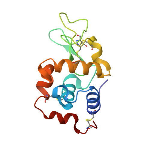
    The structures of the adducts formed upon reaction of the cytotoxic paddlewheel dirhodium complex [Rh 2 (μ-O 2 CCH 3 ) 4 ] with the model protein hen egg white lysozyme (HEWL) under different experimental conditions are reported. Results indicate that [Rh 2 (μ-O 2 CCH 3 ) 4 ] extensively reacts with HEWL:it in part breaks down, at variance with what happens in reactions with other proteins. A Rh center coordinates the side chains of Arg14 and His15. Dimeric Rh-Rh units with Rh-Rh distances between 2.3 and 2.5 Å are bound to the side chains of Asp18, Asp101, Asn93, and Lys96, while a dirhodium unit with a Rh-Rh distance of 3.2-3.4 Å binds the C-terminal carboxylate and the side chain of Lys13 at the interface between two symmetry-related molecules. An additional monometallic fragment binds the side chain of Lys33. These data, which are supported by replicated structural determinations, shed light on the reactivity of dirhodium tetracarboxylates with proteins, providing useful information for the design of new Rh-containing biomaterials with an array of potential applications in the field of catalysis or of medicinal chemistry and valuable insight into the mechanism of action of these potential anticancer agents.


  • Organizational Affiliation
    • Department of Chemical Sciences, University of Naples Federico II, 80126 Napoli, Italy.

Macromolecule Content 

  • Total Structure Weight: 15.86 kDa 
  • Atom Count: 1,282 
  • Modeled Residue Count: 129 
  • Deposited Residue Count: 129 
  • Unique protein chains: 1

Macromolecules

Find similar proteins by:|  3D Structure
Entity ID: 1
MoleculeChains  Sequence LengthOrganismDetailsImage
LysozymeA [auth AAA]129Gallus gallusMutation(s): 0 
EC: 3.2.1.17
UniProt
Find proteins for P00698 (Gallus gallus)
Explore P00698 
Go to UniProtKB:  P00698
Entity Groups
Sequence Clusters30% Identity50% Identity70% Identity90% Identity95% Identity100% Identity
UniProt GroupP00698
Sequence Annotations
Expand
Reference Sequence

Small Molecules

Ligands 5 Unique
IDChains Name / Formula / InChI Key2D Diagram3D Interactions
RH
(Subject of Investigation/LOI)

Query on RH


Download:Ideal Coordinates CCD File
K [auth AAA],
O [auth AAA]
Rhodium
Rh
OBDSITLACDGNMC-UHFFFAOYSA-N
EDO

Query on EDO


Download:Ideal Coordinates CCD File
L [auth AAA],
M [auth AAA],
N [auth AAA]
1,2-ETHANEDIOL
C2 H6 O2
LYCAIKOWRPUZTN-UHFFFAOYSA-N
NO3

Query on NO3


Download:Ideal Coordinates CCD File
C [auth AAA]
D [auth AAA]
E [auth AAA]
F [auth AAA]
G [auth AAA]
C [auth AAA],
D [auth AAA],
E [auth AAA],
F [auth AAA],
G [auth AAA],
H [auth AAA],
I [auth AAA],
J [auth AAA],
V [auth AAA],
W [auth AAA],
X [auth AAA]
NITRATE ION
N O3
NHNBFGGVMKEFGY-UHFFFAOYSA-N
ACT

Query on ACT


Download:Ideal Coordinates CCD File
P [auth AAA]
Q [auth AAA]
R [auth AAA]
S [auth AAA]
T [auth AAA]
P [auth AAA],
Q [auth AAA],
R [auth AAA],
S [auth AAA],
T [auth AAA],
U [auth AAA],
Y [auth AAA]
ACETATE ION
C2 H3 O2
QTBSBXVTEAMEQO-UHFFFAOYSA-M
NA

Query on NA


Download:Ideal Coordinates CCD File
B [auth AAA],
Z [auth AAA]
SODIUM ION
Na
FKNQFGJONOIPTF-UHFFFAOYSA-N

Experimental Data & Validation

Experimental Data

  • Method: X-RAY DIFFRACTION
  • Resolution: 1.40 Å
  • R-Value Free:  0.209 (Depositor), 0.209 (DCC) 
  • R-Value Work:  0.190 (Depositor), 0.191 (DCC) 
Space Group: P 43 21 2
Unit Cell:
Length ( Å )Angle ( ˚ )
a = 78.36α = 90
b = 78.36β = 90
c = 37.61γ = 90
Software Package:
Software NamePurpose
REFMACrefinement
MOSFLMdata reduction
MOSFLMdata scaling
PHASERphasing

Structure Validation

View Full Validation Report


Entry History 

& Funding Information

Deposition Data


Funding OrganizationLocationGrant Number
Italian Association for Cancer ResearchItaly22587

Revision History  (Full details and data files)

  • Version 1.0: 2021-02-24
    Type: Initial release
  • Version 1.1: 2024-01-31
    Changes: Data collection, Database references, Derived calculations, Refinement description
  • Version 1.2: 2024-10-23
    Changes: Structure summary