


Structure of LIMK1 Kinase domain with allosteric inhibitor TH-470
Lee, H., Yosaatmadja, Y., Burgess-Brown, N.A., von Delft, F., Arrowsmith, C.H., Edwards, A., Bountra, C., Elkins, J.M.To be published.
Experimental Data Snapshot
Starting Model: experimental
View more details
Entity ID: 1 | |||||
|---|---|---|---|---|---|
| Molecule | Chains | Sequence Length | Organism | Details | Image |
| LIM domain kinase 1 | 310 | Homo sapiens | Mutation(s): 0 Gene Names: LIMK1, LIMK EC: 2.7.11.1 | 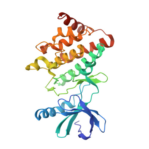 | |
UniProt & NIH Common Fund Data Resources | |||||
PHAROS: P53667 GTEx: ENSG00000106683 | |||||
Entity Groups | |||||
| Sequence Clusters | 30% Identity50% Identity70% Identity90% Identity95% Identity100% Identity | ||||
| UniProt Group | P53667 | ||||
Sequence AnnotationsExpand | |||||
Reference Sequence | |||||
| Ligands 2 Unique | |||||
|---|---|---|---|---|---|
| ID | Chains | Name / Formula / InChI Key | 2D Diagram | 3D Interactions | |
| T3B (Subject of Investigation/LOI) Download:Ideal Coordinates CCD File | E [auth A], I [auth B], J [auth C], K [auth D] | 2-(2-methylpropanoylamino)-~{N}-[2-[(phenylmethyl)-[4-(phenylsulfamoyl)phenyl]carbonyl-amino]ethyl]-1,3-thiazole-5-carboxamide C30 H31 N5 O5 S2 YVPYJSSYGACUSE-UHFFFAOYSA-N |  | ||
| EDO Download:Ideal Coordinates CCD File | F [auth A], G [auth A], H [auth A] | 1,2-ETHANEDIOL C2 H6 O2 LYCAIKOWRPUZTN-UHFFFAOYSA-N |  | ||
| Length ( Å ) | Angle ( ˚ ) |
|---|---|
| a = 84.692 | α = 90 |
| b = 83.665 | β = 91.97 |
| c = 96.513 | γ = 90 |
| Software Name | Purpose |
|---|---|
| REFMAC | refinement |
| Aimless | data scaling |
| PHASER | phasing |
| PDB_EXTRACT | data extraction |
| xia2 | data reduction |
| Funding Organization | Location | Grant Number |
|---|---|---|
| Medical Research Council (MRC, United Kingdom) | United Kingdom | -- |


