


Crystal structure of MurE from E.coli
Koekemoer, L., Steindel, M., Fairhead, M., Arrowsmith, C.H., Edwards, A.M., Bountra, C., von Delft, F., Krojer, T.To be published.
Experimental Data Snapshot
Starting Model: experimental
View more details
Entity ID: 1 | |||||
|---|---|---|---|---|---|
| Molecule | Chains | Sequence Length | Organism | Details | Image |
| UDP-N-acetylmuramoyl-L-alanyl-D-glutamate-2,6-diaminopimelate ligase | 496 | Escherichia coli K-12 | Mutation(s): 3 Gene Names: murE, b0085, JW0083 EC: 6.3.2.13 | 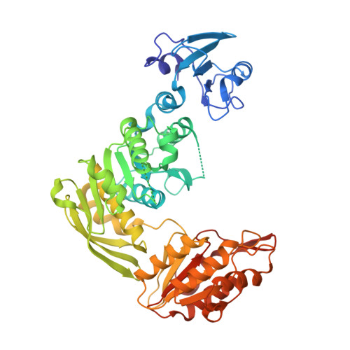 | |
UniProt | |||||
Entity Groups | |||||
| Sequence Clusters | 30% Identity50% Identity70% Identity90% Identity95% Identity100% Identity | ||||
| UniProt Group | P22188 | ||||
Sequence AnnotationsExpand | |||||
Reference Sequence | |||||
| Ligands 4 Unique | |||||
|---|---|---|---|---|---|
| ID | Chains | Name / Formula / InChI Key | 2D Diagram | 3D Interactions | |
| 9JT (Subject of Investigation/LOI) Download:Ideal Coordinates CCD File | C [auth A], G [auth B] | N-phenyl-2-selanylbenzamide C13 H11 N O Se PVPUYGNPKBMXGO-UHFFFAOYSA-N |  | ||
| CIT Download:Ideal Coordinates CCD File | F [auth A], I [auth B] | CITRIC ACID C6 H8 O7 KRKNYBCHXYNGOX-UHFFFAOYSA-N |  | ||
| EDO Download:Ideal Coordinates CCD File | J [auth B] | 1,2-ETHANEDIOL C2 H6 O2 LYCAIKOWRPUZTN-UHFFFAOYSA-N |  | ||
| IPA Download:Ideal Coordinates CCD File | D [auth A], E [auth A], H [auth B] | ISOPROPYL ALCOHOL C3 H8 O KFZMGEQAYNKOFK-UHFFFAOYSA-N |  | ||
| Length ( Å ) | Angle ( ˚ ) |
|---|---|
| a = 58.59 | α = 96.72 |
| b = 58.94 | β = 91.48 |
| c = 74.38 | γ = 104.47 |
| Software Name | Purpose |
|---|---|
| REFMAC | refinement |
| xia2 | data reduction |
| xia2 | data scaling |
| PHASER | phasing |


