7B1Z | pdb_00007b1z

Virulence-associated protein VapB from the intracellular pathogen Rhodococcus equi


Experimental Data Snapshot

  • Method: X-RAY DIFFRACTION
  • Resolution: 1.71 Å
  • R-Value Free: 
    0.182 (Depositor), 0.182 (DCC) 
  • R-Value Work: 
    0.154 (Depositor), 0.154 (DCC) 
  • R-Value Observed: 
    0.155 (Depositor) 

Starting Model: experimental
View more details

wwPDB Validation 3D Report Full Report

Validation slider image for 7B1Z

This is version 1.2 of the entry. See complete history

Literature

Conformational changes of loops highlight a potential binding site in Rhodococcus equi VapB.

Geerds, C.Haas, A.Niemann, H.H.

(2021) Acta Crystallogr F Struct Biol Commun 77: 246-253

  • DOI: https://doi.org/10.1107/S2053230X2100738X
  • Primary Citation Related Structures: 
    7B1Z

  • PubMed Abstract: 

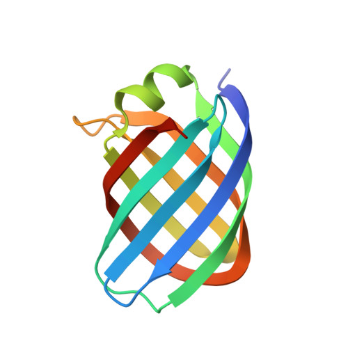
    Virulence-associated proteins (Vaps) contribute to the virulence of the pathogen Rhodococcus equi, but their mode of action has remained elusive. All Vaps share a conserved core of about 105 amino acids that folds into a compact eight-stranded antiparallel β-barrel with a unique topology. At the top of the barrel, four loops connect the eight β-strands. Previous Vap structures did not show concave surfaces that might serve as a ligand-binding site. Here, the structure of VapB in a new crystal form was determined at 1.71 Å resolution. The asymmetric unit contains two molecules. In one of them, the loop regions at the top of the barrel adopt a different conformation from other Vap structures. An outward movement of the loops results in the formation of a hydrophobic cavity that might act as a ligand-binding site. This lends further support to the hypothesis that the structural similarity between Vaps and avidins suggests a potential binding function for Vaps.


  • Organizational Affiliation
    • Department of Chemistry, Bielefeld University, Universitaetsstrasse 25, 33615 Bielefeld, Germany.

Macromolecule Content 

  • Total Structure Weight: 24.77 kDa 
  • Atom Count: 1,955 
  • Modeled Residue Count: 218 
  • Deposited Residue Count: 226 
  • Unique protein chains: 1

Macromolecules

Find similar proteins by:|  3D Structure
Entity ID: 1
MoleculeChains  Sequence LengthOrganismDetailsImage
Virulence associated protein VapB
A, B
113Prescottella equiMutation(s): 0 
Gene Names: pVAPB_vapB
Membrane Entity: Yes 
UniProt
Find proteins for B4F366 (Rhodococcus hoagii)
Explore B4F366 
Go to UniProtKB:  B4F366
Entity Groups
Sequence Clusters30% Identity50% Identity70% Identity90% Identity95% Identity100% Identity
UniProt GroupB4F366
Sequence Annotations
Expand
Reference Sequence

Experimental Data & Validation

Experimental Data

  • Method: X-RAY DIFFRACTION
  • Resolution: 1.71 Å
  • R-Value Free:  0.182 (Depositor), 0.182 (DCC) 
  • R-Value Work:  0.154 (Depositor), 0.154 (DCC) 
  • R-Value Observed: 0.155 (Depositor) 
Space Group: C 2 2 21
Unit Cell:
Length ( Å )Angle ( ˚ )
a = 56.94α = 90
b = 65.34β = 90
c = 124.26γ = 90
Software Package:
Software NamePurpose
PHENIXrefinement
XDSdata reduction
XSCALEdata scaling
PHASERphasing

Structure Validation

View Full Validation Report


Entry History 

Deposition Data

Revision History  (Full details and data files)

  • Version 1.0: 2021-07-28
    Type: Initial release
  • Version 1.1: 2021-09-01
    Changes: Database references
  • Version 1.2: 2024-01-31
    Changes: Data collection, Refinement description