7B0T | pdb_00007b0t

Crystal structure of MLLT1 YEATS domain T3 mutant in complex with benzimidazole-amide based compound 1


Experimental Data Snapshot

  • Method: X-RAY DIFFRACTION
  • Resolution: 2.05 Å
  • R-Value Free: 
    0.276 (Depositor), 0.271 (DCC) 
  • R-Value Work: 
    0.218 (Depositor) 
  • R-Value Observed: 
    0.221 (Depositor) 

Starting Model: experimental
View more details

wwPDB Validation 3D Report Full Report

Validation slider image for 7B0T

Ligand Structure Quality Assessment 


This is version 1.3 of the entry. See complete history

Literature

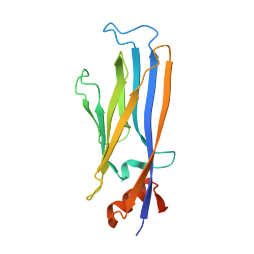
Structure and Inhibitor Binding Characterization of Oncogenic MLLT1 Mutants.

Ni, X.Londregan, A.T.Owen, D.R.Knapp, S.Chaikuad, A.

(2021) ACS Chem Biol 16: 571-578

  • DOI: https://doi.org/10.1021/acschembio.0c00960
  • Primary Citation Related Structures: 
    7B0T, 7B10

  • PubMed Abstract: 

    Dysfunction of YEATS-domain-containing MLLT1, an acetyl/acyl-lysine dependent epigenetic reader domain, has been implicated in the development of aggressive cancers. Mutations in the YEATS domain have been recently reported as a cause of MLLT1 aberrant reader function. However, the structural basis for the reported alterations in affinity for acetylated/acylated histone has remained elusive. Here, we report the crystal structures of both insertion and substitution mutants present in cancer, revealing significant conformational changes of the YEATS-domain loop 8. Structural comparison demonstrates that not only did such alteration alter the binding interface for acetylated/acylated histones, but the sequence alterations in the loop in T1 mutant may enable dimeric assembly consistent with inducing self-association behavior. Nevertheless, we show that also the MLLT1 mutants can be targeted by developed acetyllysine mimetic inhibitors with affinities similarly to wild-type. Our report provides a structural basis for the altered behaviors and a potential strategy for targeting oncogenic MLLT1 mutants.


  • Organizational Affiliation
    • Structural Genomics Consortium and Buchmann Institute for Molecular Life Sciences, Goethe University Frankfurt, Max-von-Laue-Straße 15, 60438 Frankfurt am Main, Germany.

Macromolecule Content 

  • Total Structure Weight: 18.83 kDa 
  • Atom Count: 1,183 
  • Modeled Residue Count: 138 
  • Deposited Residue Count: 152 
  • Unique protein chains: 1

Macromolecules

Find similar proteins by:|  3D Structure
Entity ID: 1
MoleculeChains  Sequence LengthOrganismDetailsImage
Protein ENL152Homo sapiensMutation(s): 1 
Gene Names: MLLT1ENLLTG19YEATS1
UniProt & NIH Common Fund Data Resources
Find proteins for Q03111 (Homo sapiens)
Explore Q03111 
Go to UniProtKB:  Q03111
PHAROS:  Q03111
GTEx:  ENSG00000130382 
Entity Groups
Sequence Clusters30% Identity50% Identity70% Identity90% Identity95% Identity100% Identity
UniProt GroupQ03111
Sequence Annotations
Expand
Reference Sequence

Experimental Data & Validation

Experimental Data

  • Method: X-RAY DIFFRACTION
  • Resolution: 2.05 Å
  • R-Value Free:  0.276 (Depositor), 0.271 (DCC) 
  • R-Value Work:  0.218 (Depositor) 
  • R-Value Observed: 0.221 (Depositor) 
Space Group: P 43 21 2
Unit Cell:
Length ( Å )Angle ( ˚ )
a = 48.739α = 90
b = 48.739β = 90
c = 131.995γ = 90
Software Package:
Software NamePurpose
Aimlessdata scaling
REFMACrefinement
PDB_EXTRACTdata extraction
XDSdata reduction
MLPHAREphasing

Structure Validation

View Full Validation Report


Ligand Structure Quality Assessment 


Entry History 

Deposition Data

Revision History  (Full details and data files)

  • Version 1.0: 2021-02-17
    Type: Initial release
  • Version 1.1: 2021-04-14
    Changes: Database references
  • Version 1.2: 2021-04-28
    Changes: Database references
  • Version 1.3: 2024-01-31
    Changes: Data collection, Database references, Refinement description