7AZT | pdb_00007azt

X-ray crystallographic structure of (6-4)photolyase from Drosophila melanogaster at room temperature


Experimental Data Snapshot

  • Method: X-RAY DIFFRACTION
  • Resolution: 2.27 Å
  • R-Value Free: 
    0.208 (Depositor), 0.218 (DCC) 
  • R-Value Work: 
    0.172 (Depositor), 0.180 (DCC) 
  • R-Value Observed: 
    0.174 (Depositor) 

Starting Model: experimental
View more details

wwPDB Validation 3D Report Full Report

Validation slider image for 7AZT

Ligand Structure Quality Assessment 


This is version 1.1 of the entry. See complete history

Literature

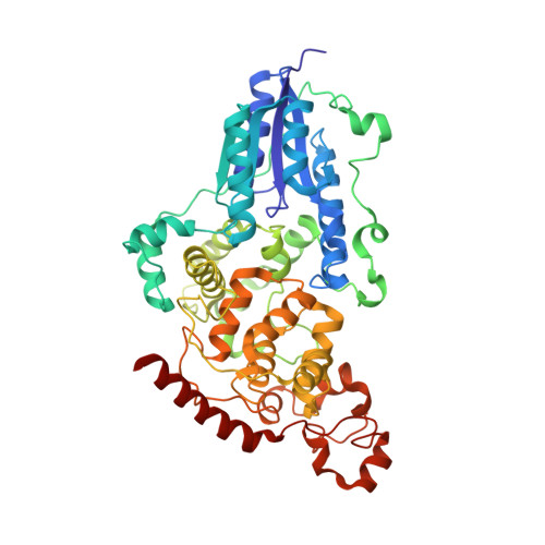
The three-dimensional structure of Drosophila melanogaster (6-4) photolyase at room temperature.

Cellini, A.Yuan Wahlgren, W.Henry, L.Pandey, S.Ghosh, S.Castillon, L.Claesson, E.Takala, H.Kubel, J.Nimmrich, A.Kuznetsova, V.Nango, E.Iwata, S.Owada, S.Stojkovic, E.A.Schmidt, M.Ihalainen, J.A.Westenhoff, S.

(2021) Acta Crystallogr D Struct Biol 77: 1001-1009

  • DOI: https://doi.org/10.1107/S2059798321005830
  • Primary Citation Related Structures: 
    7AYV, 7AZT

  • PubMed Abstract: 

    (6-4) photolyases are flavoproteins that belong to the photolyase/cryptochrome family. Their function is to repair DNA lesions using visible light. Here, crystal structures of Drosophila melanogaster (6-4) photolyase [Dm(6-4)photolyase] at room and cryogenic temperatures are reported. The room-temperature structure was solved to 2.27 Å resolution and was obtained by serial femtosecond crystallography (SFX) using an X-ray free-electron laser. The crystallization and preparation conditions are also reported. The cryogenic structure was solved to 1.79 Å resolution using conventional X-ray crystallography. The structures agree with each other, indicating that the structural information obtained from crystallography at cryogenic temperature also applies at room temperature. Furthermore, UV-Vis absorption spectroscopy confirms that Dm(6-4)photolyase is photoactive in the crystals, giving a green light to time-resolved SFX studies on the protein, which can reveal the structural mechanism of the photoactivated protein in DNA repair.


  • Organizational Affiliation
    • Department of Chemistry and Molecular Biology, University of Gothenburg, Box 462, 405 30 Gothenburg, Sweden.

Macromolecule Content 

  • Total Structure Weight: 59.1 kDa 
  • Atom Count: 4,168 
  • Modeled Residue Count: 502 
  • Deposited Residue Count: 502 
  • Unique protein chains: 1

Macromolecules

Find similar proteins by:|  3D Structure
Entity ID: 1
MoleculeChains  Sequence LengthOrganismDetailsImage
RE11660p502Drosophila melanogasterMutation(s): 0 
Gene Names: phr6-4CG2488
UniProt
Find proteins for Q8SXK5 (Drosophila melanogaster)
Explore Q8SXK5 
Go to UniProtKB:  Q8SXK5
Entity Groups
Sequence Clusters30% Identity50% Identity70% Identity90% Identity95% Identity100% Identity
UniProt GroupQ8SXK5
Sequence Annotations
Expand
Reference Sequence

Small Molecules

Ligands 1 Unique
IDChains Name / Formula / InChI Key2D Diagram3D Interactions
FAD
(Subject of Investigation/LOI)

Query on FAD


Download:Ideal Coordinates CCD File
B [auth A]FLAVIN-ADENINE DINUCLEOTIDE
C27 H33 N9 O15 P2
VWWQXMAJTJZDQX-UYBVJOGSSA-N

Experimental Data & Validation

Experimental Data

  • Method: X-RAY DIFFRACTION
  • Resolution: 2.27 Å
  • R-Value Free:  0.208 (Depositor), 0.218 (DCC) 
  • R-Value Work:  0.172 (Depositor), 0.180 (DCC) 
  • R-Value Observed: 0.174 (Depositor) 
Space Group: P 21 21 21
Unit Cell:
Length ( Å )Angle ( ˚ )
a = 61.63α = 90
b = 91.64β = 90
c = 115.97γ = 90
Software Package:
Software NamePurpose
PHENIXrefinement
CrystFELdata reduction
CrystFELdata scaling
PHASERphasing

Structure Validation

View Full Validation Report


Ligand Structure Quality Assessment 


Entry History 

& Funding Information

Deposition Data


Funding OrganizationLocationGrant Number
Swedish Research CouncilSweden--

Revision History  (Full details and data files)

  • Version 1.0: 2021-08-18
    Type: Initial release
  • Version 1.1: 2024-01-31
    Changes: Data collection, Refinement description