7AZP | pdb_00007azp

Structure of the human mitochondrial HSPD1 single ring


Experimental Data Snapshot

  • Method: ELECTRON MICROSCOPY
  • Resolution: 3.50 Å
  • Aggregation State: PARTICLE 
  • Reconstruction Method: SINGLE PARTICLE 

Starting Models: experimental
View more details

wwPDB Validation 3D Report Full Report

Validation slider image for 7AZP

This is version 1.1 of the entry. See complete history

Literature

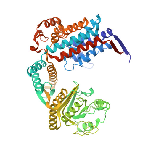
Cryo-EM structure of human mitochondrial HSPD1.

Klebl, D.P.Feasey, M.C.Hesketh, E.L.Ranson, N.A.Wurdak, H.Sobott, F.Bon, R.S.Muench, S.P.

(2021) iScience 24: 102022-102022

  • DOI: https://doi.org/10.1016/j.isci.2020.102022
  • Primary Citation Related Structures: 
    7AZP

  • PubMed Abstract: 

    Chaperonins play an important role in folding newly synthesized or translocated proteins in all organisms. The bacterial chaperonin GroEL has served as a model system for the understanding of these proteins. In comparison, its human homolog, known as mitochondrial heat shock protein family member D1 (HSPD1) is poorly understood. Here, we present the structure of HSPD1 in the apo state determined by cryo-electron microscopy (cryo-EM). Unlike GroEL, HSPD1 forms mostly single ring assemblies in the absence of co-chaperonin (HSPE1). Comparison with GroEL shows a rotation and increased flexibility of the apical domain. Together with published structures of the HSPD1/HSPE1 co-chaperonin complex, this work gives insight into the structural changes that occur during the catalytic cycle. This new understanding of HSPD1 structure and its rearrangements upon complex formation may provide new insights for the development of HSPD1-targeting treatments against a diverse range of diseases including glioblastoma.


  • Organizational Affiliation
    • School of Biomedical Sciences, Faculty of Biological Sciences & Astbury Centre for Structural and Molecular Biology, University of Leeds, Leeds LS2 9JT, UK.

Macromolecule Content 

  • Total Structure Weight: 408.15 kDa 
  • Atom Count: 27,391 
  • Modeled Residue Count: 3,675 
  • Deposited Residue Count: 3,850 
  • Unique protein chains: 1

Macromolecules

Find similar proteins by:|  3D Structure
Entity ID: 1
MoleculeChains  Sequence LengthOrganismDetailsImage
60 kDa heat shock protein, mitochondrial
A, B, C, D, E
A, B, C, D, E, F, G
550Homo sapiensMutation(s): 0 
Gene Names: HSPD1HSP60
EC: 5.6.1.7
UniProt & NIH Common Fund Data Resources
Find proteins for P10809 (Homo sapiens)
Explore P10809 
Go to UniProtKB:  P10809
PHAROS:  P10809
GTEx:  ENSG00000144381 
Entity Groups
Sequence Clusters30% Identity50% Identity70% Identity90% Identity95% Identity100% Identity
UniProt GroupP10809
Sequence Annotations
Expand
Reference Sequence

Experimental Data & Validation

Experimental Data

  • Method: ELECTRON MICROSCOPY
  • Resolution: 3.50 Å
  • Aggregation State: PARTICLE 
  • Reconstruction Method: SINGLE PARTICLE 
EM Software:
TaskSoftware PackageVersion
MODEL REFINEMENTPHENIX
RECONSTRUCTIONRELION

Structure Validation

View Full Validation Report


Entry History 

& Funding Information

Deposition Data


Funding OrganizationLocationGrant Number
Biotechnology and Biological Sciences Research Council (BBSRC)United KingdomBB/P026397/1
Wellcome TrustUnited Kingdom108466/Z/15/Z

Revision History  (Full details and data files)

  • Version 1.0: 2021-02-10
    Type: Initial release
  • Version 1.1: 2024-05-01
    Changes: Data collection, Database references, Refinement description