7AX2 | pdb_00007ax2

Crystal structure of the computationally designed Scone-E protein co-crystallized with STA, form b


Experimental Data Snapshot

  • Method: X-RAY DIFFRACTION
  • Resolution: 2.10 Å
  • R-Value Free: 
    0.206 (Depositor), 0.231 (DCC) 
  • R-Value Work: 
    0.178 (Depositor) 
  • R-Value Observed: 
    0.180 (Depositor) 

Starting Model: experimental
View more details

wwPDB Validation 3D Report Full Report

Validation slider image for 7AX2

Ligand Structure Quality Assessment 


This is version 2.2 of the entry. See complete history

Literature

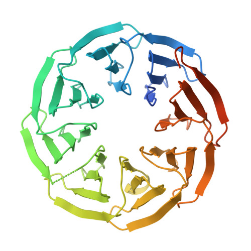
Crystal structures of Scone: pseudosymmetric folding of a symmetric designer protein.

Mylemans, B.Killian, T.Vandebroek, L.Van Meervelt, L.Tame, J.R.H.Parac-Vogt, T.N.Voet, A.R.D.

(2021) Acta Crystallogr D Struct Biol 77: 933-942

  • DOI: https://doi.org/10.1107/S2059798321005787
  • Primary Citation Related Structures: 
    7AWY, 7AWZ, 7AX0, 7AX2

  • PubMed Abstract: 

    Recent years have seen an increase in the development of computational proteins, including symmetric ones. A ninefold-symmetric β-propeller protein named Cake has recently been developed. Here, attempts were made to further engineer this protein into a threefold-symmetric nine-bladed propeller using computational design. Two nine-bladed propeller proteins were designed, named Scone-E and Scone-R. Crystallography, however, revealed the structure of both designs to adopt an eightfold conformation with distorted termini, leading to a pseudo-symmetric protein. One of the proteins could only be crystallized upon the addition of a polyoxometalate, highlighting the usefulness of these molecules as crystallization additives.


  • Organizational Affiliation
    • Laboratory of Biomolecular Modelling and Design, Department of Chemistry, KU Leuven, Celestijnenlaan 200G, 3001 Leuven, Vlaams Brabant, Belgium.

Macromolecule Content 

  • Total Structure Weight: 45.69 kDa 
  • Atom Count: 2,534 
  • Modeled Residue Count: 311 
  • Deposited Residue Count: 358 
  • Unique protein chains: 1

Macromolecules

Find similar proteins by:|  3D Structure
Entity ID: 1
MoleculeChains  Sequence LengthOrganismDetailsImage
Scone-E358synthetic constructMutation(s): 0 

Experimental Data & Validation

Experimental Data

  • Method: X-RAY DIFFRACTION
  • Resolution: 2.10 Å
  • R-Value Free:  0.206 (Depositor), 0.231 (DCC) 
  • R-Value Work:  0.178 (Depositor) 
  • R-Value Observed: 0.180 (Depositor) 
Space Group: I 41 2 2
Unit Cell:
Length ( Å )Angle ( ˚ )
a = 133.517α = 90
b = 133.517β = 90
c = 70.093γ = 90
Software Package:
Software NamePurpose
PHENIXrefinement
DIALSdata reduction
Aimlessdata scaling
PHASERphasing

Structure Validation

View Full Validation Report


Ligand Structure Quality Assessment 


Entry History 

& Funding Information

Deposition Data


Funding OrganizationLocationGrant Number
Research Foundation - Flanders (FWO)BelgiumGBM-D3229-ASP/17
Research Foundation - Flanders (FWO)BelgiumG0E4717N
Research Foundation - Flanders (FWO)BelgiumG051917N
Research Foundation - Flanders (FWO)BelgiumG0F9316N

Revision History  (Full details and data files)

  • Version 1.0: 2021-01-13
    Type: Initial release
  • Version 2.0: 2021-06-30
    Changes: Database references, Non-polymer description
  • Version 2.1: 2021-07-14
    Changes: Database references
  • Version 2.2: 2024-01-31
    Changes: Data collection, Database references, Refinement description