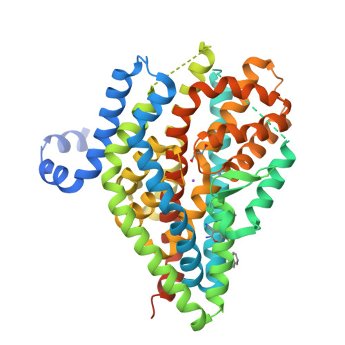
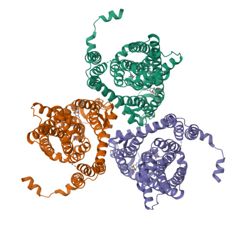
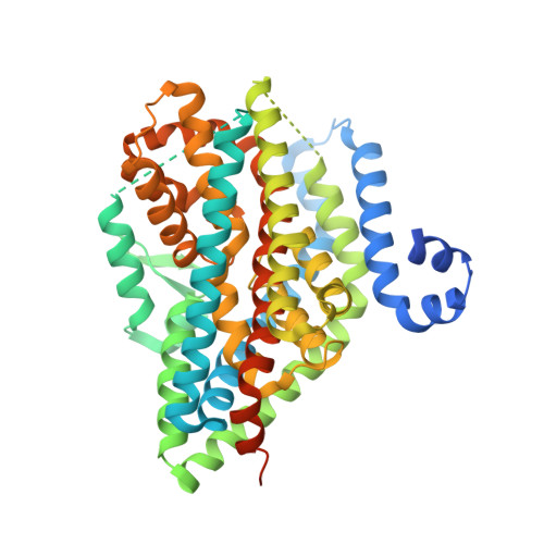
7AWM | pdb_00007awm

Structure of the thermostabilized EAAT1 cryst mutant in complex with L-ASP, three sodium ions and the allosteric inhibitor UCPH101


Experimental Data Snapshot

  • Method: X-RAY DIFFRACTION
  • Resolution: 3.25 Å
  • R-Value Free: 
    0.238 (Depositor), 0.270 (DCC) 
  • R-Value Work: 
    0.219 (Depositor), 0.230 (DCC) 
  • R-Value Observed: 
    0.220 (Depositor) 

Starting Model: experimental
View more details

wwPDB Validation   3D Report Full Report


Ligand Structure Quality Assessment 

Created with Raphaël 2.3.0Worse 01 BetterLigand structure goodness of fit to experimental dataBest fitted 6Z6Click on this verticalbar to view details

This is version 1.4 of the entry. See complete history


Literature

The ion-coupling mechanism of human excitatory amino acid transporters.

Canul-Tec, J.C.Kumar, A.Dhenin, J.Assal, R.Legrand, P.Rey, M.Chamot-Rooke, J.Reyes, N.

(2022) EMBO J 41: e108341-e108341

  • DOI: https://doi.org/10.15252/embj.2021108341
  • Primary Citation of Related Structures:  
    7AWL, 7AWM, 7AWN, 7AWP, 7AWQ, 7NPW

  • PubMed Abstract: 

    Excitatory amino acid transporters (EAATs) maintain glutamate gradients in the brain essential for neurotransmission and to prevent neuronal death. They use ionic gradients as energy source and co-transport transmitter into the cytoplasm with Na + and H + , while counter-transporting K + to re-initiate the transport cycle. However, the molecular mechanisms underlying ion-coupled transport remain incompletely understood. Here, we present 3D X-ray crystallographic and cryo-EM structures, as well as thermodynamic analysis of human EAAT1 in different ion bound conformations, including elusive counter-transport ion bound states. Binding energies of Na + and H + , and unexpectedly Ca 2+ , are coupled to neurotransmitter binding. Ca 2+ competes for a conserved Na + site, suggesting a regulatory role for Ca 2+ in glutamate transport at the synapse, while H + binds to a conserved glutamate residue stabilizing substrate occlusion. The counter-transported ion binding site overlaps with that of glutamate, revealing the K + -based mechanism to exclude the transmitter during the transport cycle and to prevent its neurotoxic release on the extracellular side.


  • Organizational Affiliation

    Membrane Protein Mechanisms Unit, Institut Pasteur, Paris, France.


Macromolecules
Find similar proteins by:  (by identity cutoff)  |  3D Structure
Entity ID: 1
MoleculeChains Sequence LengthOrganismDetailsImage
Excitatory amino acid transporter 1,Neutral amino acid transporter B(0),Excitatory amino acid transporter 1522Homo sapiensMutation(s): 72 
Gene Names: SLC1A3EAAT1GLASTGLAST1SLC1A5ASCT2M7V1RDRRDRC
Membrane Entity: Yes 
UniProt & NIH Common Fund Data Resources
Find proteins for Q15758 (Homo sapiens)
Explore Q15758 
Go to UniProtKB:  Q15758
PHAROS:  Q15758
GTEx:  ENSG00000105281 
Find proteins for P43003 (Homo sapiens)
Explore P43003 
Go to UniProtKB:  P43003
PHAROS:  P43003
GTEx:  ENSG00000079215 
Entity Groups  
Sequence Clusters30% Identity50% Identity70% Identity90% Identity95% Identity100% Identity
UniProt GroupsP43003Q15758
Sequence Annotations
Expand
  • Reference Sequence
Small Molecules
Ligands 4 Unique
IDChains Name / Formula / InChI Key2D Diagram3D Interactions
6Z6 (Subject of Investigation/LOI)
Query on 6Z6

Download Ideal Coordinates CCD File 
F [auth A]2-Amino-5,6,7,8-tetrahydro-4-(4-methoxyphenyl)-7-(naphthalen-1-yl)-5-oxo-4H-chromene-3-carbonitrile
C27 H22 N2 O3
YBMGNDPBARCLFT-YDONVPIESA-N
BA
Query on BA

Download Ideal Coordinates CCD File 
G [auth A]BARIUM ION
Ba
XDFCIPNJCBUZJN-UHFFFAOYSA-N
ASP
Query on ASP

Download Ideal Coordinates CCD File 
E [auth A]ASPARTIC ACID
C4 H7 N O4
CKLJMWTZIZZHCS-REOHCLBHSA-N
NA
Query on NA

Download Ideal Coordinates CCD File 
B [auth A],
C [auth A],
D [auth A]
SODIUM ION
Na
FKNQFGJONOIPTF-UHFFFAOYSA-N
Experimental Data & Validation

Experimental Data

  • Method: X-RAY DIFFRACTION
  • Resolution: 3.25 Å
  • R-Value Free:  0.238 (Depositor), 0.270 (DCC) 
  • R-Value Work:  0.219 (Depositor), 0.230 (DCC) 
  • R-Value Observed: 0.220 (Depositor) 
Space Group: P 63
Unit Cell:
Length ( Å )Angle ( ˚ )
a = 122.882α = 90
b = 122.882β = 90
c = 90.032γ = 120
Software Package:
Software NamePurpose
BUSTERrefinement
XDSdata reduction
Aimlessdata scaling
PHASERphasing
PDB_EXTRACTdata extraction

Structure Validation

View Full Validation Report


Ligand Structure Quality Assessment 

Created with Raphaël 2.3.0Worse 01 BetterLigand structure goodness of fit to experimental dataBest fitted 6Z6Click on this verticalbar to view details

Entry History & Funding Information

Deposition Data


Funding OrganizationLocationGrant Number
European Research Council (ERC)France309657

Revision History  (Full details and data files)

  • Version 1.0: 2021-10-13
    Type: Initial release
  • Version 1.1: 2021-12-01
    Changes: Database references
  • Version 1.2: 2022-01-12
    Changes: Database references
  • Version 1.3: 2022-01-19
    Changes: Database references
  • Version 1.4: 2024-01-31
    Changes: Data collection, Refinement description