7AS6

2.0 angstrom structure of plant Extended Synaptotagmin 1, C2A domain


Experimental Data Snapshot

  • Method: X-RAY DIFFRACTION
  • Resolution: 2.00 Å
  • R-Value Free: 0.211 
  • R-Value Work: 0.186 
  • R-Value Observed: 0.187 

wwPDB Validation   3D Report Full Report


This is version 1.2 of the entry. See complete history


Literature

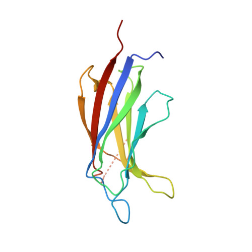
The structure and flexibility analysis of the Arabidopsis synaptotagmin 1 reveal the basis of its regulation at membrane contact sites.

Benavente, J.L.Siliqi, D.Infantes, L.Lagartera, L.Mills, A.Gago, F.Ruiz-Lopez, N.Botella, M.A.Sanchez-Barrena, M.J.Albert, A.

(2021) Life Sci Alliance 4

  • DOI: https://doi.org/10.26508/lsa.202101152
  • Primary Citation of Related Structures:  
    7AS6, 7ATP

  • PubMed Abstract: 

    Non-vesicular lipid transfer at ER and plasma membrane (PM) contact sites (CS) is crucial for the maintenance of membrane lipid homeostasis. Extended synaptotagmins (E-Syts) play a central role in this process as they act as molecular tethers of ER and PM and as lipid transfer proteins between these organelles. E-Syts are proteins constitutively anchored to the ER through an N-terminal hydrophobic segment and bind the PM via a variable number of C-terminal C2 domains. Synaptotagmins (SYTs) are the plant orthologous of E-Syts and regulate the ER-PM communication in response to abiotic stress. Combining different structural and biochemical techniques, we demonstrate that the binding of SYT1 to lipids occurs through a Ca 2+ -dependent lipid-binding site and by a site for phosphorylated forms of phosphatidylinositol, thus integrating two different molecular signals in response to stress. In addition, we show that SYT1 displays three highly flexible hinge points that provide conformational freedom to facilitate lipid extraction, protein loading, and subsequent transfer between PM and ER.


  • Organizational Affiliation

    Instituto de Química Física "Rocasolano," Consejo Superior de Investigaciones Científicas (CSIC), Madrid, Spain.


Macromolecules
Find similar proteins by:  (by identity cutoff)  |  3D Structure
Entity ID: 1
MoleculeChains Sequence LengthOrganismDetailsImage
Synaptotagmin-1145Arabidopsis thalianaMutation(s): 0 
Gene Names: SYT1SYTAAt2g20990F26H11.25F5H14.5
Membrane Entity: Yes 
UniProt
Find proteins for Q9SKR2 (Arabidopsis thaliana)
Explore Q9SKR2 
Go to UniProtKB:  Q9SKR2
Entity Groups  
Sequence Clusters30% Identity50% Identity70% Identity90% Identity95% Identity100% Identity
UniProt GroupQ9SKR2
Sequence Annotations
Expand
  • Reference Sequence
Small Molecules
Ligands 4 Unique
IDChains Name / Formula / InChI Key2D Diagram3D Interactions
CD
Query on CD

Download Ideal Coordinates CCD File 
B [auth A],
C [auth A]
CADMIUM ION
Cd
WLZRMCYVCSSEQC-UHFFFAOYSA-N
EDO
Query on EDO

Download Ideal Coordinates CCD File 
G [auth A],
H [auth A]
1,2-ETHANEDIOL
C2 H6 O2
LYCAIKOWRPUZTN-UHFFFAOYSA-N
NI
Query on NI

Download Ideal Coordinates CCD File 
D [auth A],
E [auth A]
NICKEL (II) ION
Ni
VEQPNABPJHWNSG-UHFFFAOYSA-N
CL
Query on CL

Download Ideal Coordinates CCD File 
F [auth A]CHLORIDE ION
Cl
VEXZGXHMUGYJMC-UHFFFAOYSA-M
Experimental Data & Validation

Experimental Data

  • Method: X-RAY DIFFRACTION
  • Resolution: 2.00 Å
  • R-Value Free: 0.211 
  • R-Value Work: 0.186 
  • R-Value Observed: 0.187 
  • Space Group: H 3
Unit Cell:
Length ( Å )Angle ( ˚ )
a = 70.937α = 90
b = 70.937β = 90
c = 116.309γ = 120
Software Package:
Software NamePurpose
PHENIXrefinement
XDSdata reduction
XDSdata scaling
PHASERphasing

Structure Validation

View Full Validation Report


Entry History 

Deposition Data

Revision History  (Full details and data files)

  • Version 1.0: 2021-08-18
    Type: Initial release
  • Version 1.1: 2021-10-06
    Changes: Data collection, Database references, Structure summary
  • Version 1.2: 2024-01-31
    Changes: Data collection, Refinement description