7AOV | pdb_00007aov

Crystal Structure of a TRPM2 Domain


Experimental Data Snapshot

  • Method: X-RAY DIFFRACTION
  • Resolution: 2.00 Å
  • R-Value Free: 
    0.231 (Depositor), 0.235 (DCC) 
  • R-Value Work: 
    0.188 (Depositor), 0.191 (DCC) 
  • R-Value Observed: 
    0.190 (Depositor) 

wwPDB Validation 3D Report Full Report

Validation slider image for 7AOV

This is version 1.2 of the entry. See complete history

Literature

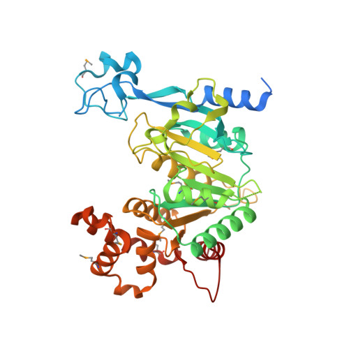
The crystal structure of TRPM2 MHR1/2 domain reveals a conserved Zn 2+ -binding domain essential for structural integrity and channel activity.

Sander, S.Pick, J.Gattkowski, E.Fliegert, R.Tidow, H.

(2022) Protein Sci 31: e4320-e4320

  • DOI: https://doi.org/10.1002/pro.4320
  • Primary Citation Related Structures: 
    7AOV

  • PubMed Abstract: 

    Transient receptor potential melastatin 2 (TRPM2) is a Ca 2+ -permeable, nonselective cation channel involved in diverse physiological processes such as immune response, apoptosis, and body temperature sensing. TRPM2 is activated by ADP-ribose (ADPR) and 2'-deoxy-ADPR in a Ca 2+ -dependent manner. While two distinct binding sites exist for ADPR that exert different functions dependent on the species, the involvement of either binding site regarding the superagonistic effect of 2'-deoxy-ADPR is not clear yet. Here, we report the crystal structure of the MHR1/2 domain of TRPM2 from zebrafish (Danio rerio), and show that both ligands bind to this domain and activate the channel. We identified a so far unrecognized Zn 2+ -binding domain that was not resolved in previous cryo-EM structures and that is conserved in most TRPM channels. In combination with patch clamp experiments we comprehensively characterize the effect of the Zn 2+ -binding domain on TRPM2 activation. Our results provide insight into a conserved motif essential for structural integrity and channel activity.


  • Organizational Affiliation
    • Hamburg Advanced Research Centre for Bioorganic Chemistry (HARBOR) & Department of Chemistry, Institute for Biochemistry and Molecular Biology, University of Hamburg, Hamburg, Germany.

Macromolecule Content 

  • Total Structure Weight: 96.13 kDa 
  • Atom Count: 6,370 
  • Modeled Residue Count: 754 
  • Deposited Residue Count: 848 
  • Unique protein chains: 1

Macromolecules

Find similar proteins by:|  3D Structure
Entity ID: 1
MoleculeChains  Sequence LengthOrganismDetailsImage
Transient receptor potential cation channel subfamily M member 2
A, B
424Danio rerioMutation(s): 0 
Gene Names: trpm2
UniProt
Find proteins for A0A0R4IMY7 (Danio rerio)
Explore A0A0R4IMY7 
Go to UniProtKB:  A0A0R4IMY7
Entity Groups
Sequence Clusters30% Identity50% Identity70% Identity90% Identity95% Identity100% Identity
UniProt GroupA0A0R4IMY7
Sequence Annotations
Expand
Reference Sequence

Small Molecules

Modified Residues  1 Unique
IDChains TypeFormula2D DiagramParent
MSE
Query on MSE
A, B
L-PEPTIDE LINKINGC5 H11 N O2 SeMET

Experimental Data & Validation

Experimental Data

  • Method: X-RAY DIFFRACTION
  • Resolution: 2.00 Å
  • R-Value Free:  0.231 (Depositor), 0.235 (DCC) 
  • R-Value Work:  0.188 (Depositor), 0.191 (DCC) 
  • R-Value Observed: 0.190 (Depositor) 
Space Group: P 1 21 1
Unit Cell:
Length ( Å )Angle ( ˚ )
a = 47.76α = 90
b = 188.128β = 102.659
c = 47.773γ = 90
Software Package:
Software NamePurpose
PHENIXrefinement
XDSdata reduction
Aimlessdata scaling
SHELXCDphasing
ARP/wARPmodel building

Structure Validation

View Full Validation Report


Entry History 

& Funding Information

Deposition Data


Funding OrganizationLocationGrant Number
German Research Foundation (DFG)GermanySFB 1328

Revision History  (Full details and data files)

  • Version 1.0: 2022-03-02
    Type: Initial release
  • Version 1.1: 2022-08-17
    Changes: Database references
  • Version 1.2: 2024-11-06
    Changes: Data collection, Structure summary