7AML | pdb_00007aml

RET/GDNF/GFRa1 extracellular complex Cryo-EM structure


Experimental Data Snapshot

  • Method: ELECTRON MICROSCOPY
  • Resolution: 3.50 Å
  • Aggregation State: PARTICLE 
  • Reconstruction Method: SINGLE PARTICLE 

Starting Model: experimental
View more details

wwPDB Validation 3D Report Full Report

Validation slider image for 7AML

This is version 1.3 of the entry. See complete history. 

Literature

A two-site flexible clamp mechanism for RET-GDNF-GFR alpha 1 assembly reveals both conformational adaptation and strict geometric spacing.

Adams, S.E., Purkiss, A.G., Knowles, P.P., Nans, A., Briggs, D.C., Borg, A., Earl, C.P., Goodman, K.M., Nawrotek, A., Borg, A.J., McIntosh, P.B., Houghton, F.M., Kjaer, S., McDonald, N.Q.

(2021) Structure 29: 694

  • DOI: https://doi.org/10.1016/j.str.2020.12.012
  • Primary Citation Related Structures: 
    7AB8, 7AMK, 7AML

  • PubMed Abstract: 

    RET receptor tyrosine kinase plays vital developmental and neuroprotective roles in metazoans. GDNF family ligands (GFLs) when bound to cognate GFRα co-receptors recognize and activate RET stimulating its cytoplasmic kinase function. The principles for RET ligand-co-receptor recognition are incompletely understood. Here, we report a crystal structure of the cadherin-like module (CLD1-4) from zebrafish RET revealing interdomain flexibility between CLD2 and CLD3. Comparison with a cryo-electron microscopy structure of a ligand-engaged zebrafish RET ECD -GDNF-GFRα1a complex indicates conformational changes within a clade-specific CLD3 loop adjacent to the co-receptor. Our observations indicate that RET is a molecular clamp with a flexible calcium-dependent arm that adapts to different GFRα co-receptors, while its rigid arm recognizes a GFL dimer to align both membrane-proximal cysteine-rich domains. We also visualize linear arrays of RET ECD -GDNF-GFRα1a suggesting that a conserved contact stabilizes higher-order species. Our study reveals that ligand-co-receptor recognition by RET involves both receptor plasticity and strict spacing of receptor dimers by GFL ligands.


  • Organizational Affiliation: 
    • Signalling and Structural Biology Laboratory, Francis Crick Institute, NW1 1AT London, UK.

Macromolecule Content 

  • Total Structure Weight: 245.54 kDa 
  • Atom Count: 16,018 
  • Modeled Residue Count: 1,996 
  • Deposited Residue Count: 2,138 
  • Unique protein chains: 3

Macromolecules

Find similar proteins by:|  3D Structure
Entity ID: 1
MoleculeChains  Sequence LengthOrganismDetailsImage
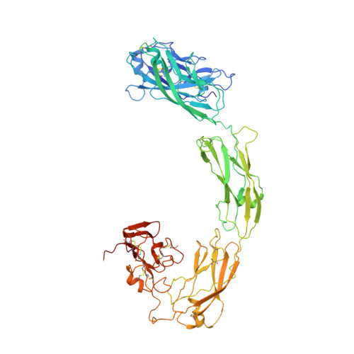
Proto-oncogene tyrosine-protein kinase receptor RetA,
B [auth D]
615Danio rerioMutation(s): 0 
Gene Names: ret, ret1
EC: 2.7.10.1
UniProt
Find proteins for A8E7C6 (Danio rerio)
Explore A8E7C6 
Go to UniProtKB:  A8E7C6
Entity Groups
Sequence Clusters30% Identity50% Identity70% Identity90% Identity95% Identity100% Identity
UniProt GroupA8E7C6
Glycosylation
Glycosylation Sites: 7
Sequence Annotations
Expand
Reference Sequence
Find similar proteins by:|  3D Structure
Entity ID: 2
MoleculeChains  Sequence LengthOrganismDetailsImage
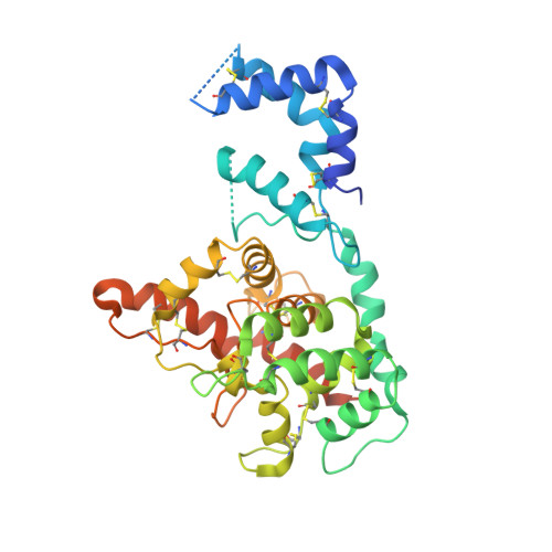
GDNF family receptor alphaC [auth B],
E
353Danio rerioMutation(s): 0 
Gene Names: gfra1a, gfralpha1a
UniProt
Find proteins for Q98TT9 (Danio rerio)
Explore Q98TT9 
Go to UniProtKB:  Q98TT9
Entity Groups
Sequence Clusters30% Identity50% Identity70% Identity90% Identity95% Identity100% Identity
UniProt GroupQ98TT9
Sequence Annotations
Expand
Reference Sequence
Find similar proteins by:|  3D Structure
Entity ID: 3
MoleculeChains  Sequence LengthOrganismDetailsImage
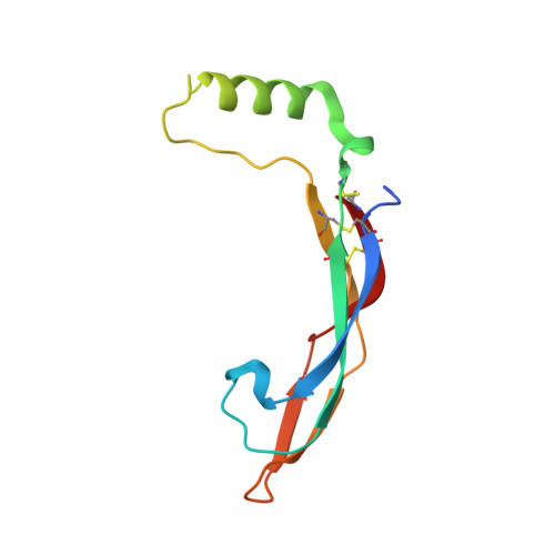
Glial cell line-derived neurotrophic factorD [auth C],
F
101Danio rerioMutation(s): 0 
Gene Names: gdnf
UniProt
Find proteins for Q98TU0 (Danio rerio)
Explore Q98TU0 
Go to UniProtKB:  Q98TU0
Entity Groups
Sequence Clusters30% Identity50% Identity70% Identity90% Identity95% Identity100% Identity
UniProt GroupQ98TU0
Glycosylation
Glycosylation Sites: 1
Sequence Annotations
Expand
Reference Sequence

Small Molecules

Ligands 2 Unique
IDChains Name / Formula / InChI Key2D Diagram3D Interactions
NAG

Query on NAG


Download:Ideal Coordinates CCD File
AA [auth D]
BA [auth D]
CA [auth C]
DA [auth F]
K [auth A]
AA [auth D],
BA [auth D],
CA [auth C],
DA [auth F],
K [auth A],
L [auth A],
M [auth A],
N [auth A],
O [auth A],
P [auth A],
Q [auth A],
V [auth D],
W [auth D],
X [auth D],
Y [auth D],
Z [auth D]
2-acetamido-2-deoxy-beta-D-glucopyranose
C8 H15 N O6
OVRNDRQMDRJTHS-FMDGEEDCSA-N
CA

Query on CA


Download:Ideal Coordinates CCD File
G [auth A]
H [auth A]
I [auth A]
J [auth A]
R [auth D]
G [auth A],
H [auth A],
I [auth A],
J [auth A],
R [auth D],
S [auth D],
T [auth D],
U [auth D]
CALCIUM ION
Ca
BHPQYMZQTOCNFJ-UHFFFAOYSA-N

Experimental Data & Validation

Experimental Data

  • Method: ELECTRON MICROSCOPY
  • Resolution: 3.50 Å
  • Aggregation State: PARTICLE 
  • Reconstruction Method: SINGLE PARTICLE 
EM Software:
TaskSoftware PackageVersion
RECONSTRUCTIONcryoSPARC2
MODEL REFINEMENTPHENIX1.18.2
MODEL REFINEMENTCoot

Structure Validation

View Full Validation Report


Entry History 

& Funding Information

Deposition Data


Funding OrganizationLocationGrant Number
The Francis Crick InstituteUnited Kingdom10115

Revision History  (Full details and data files)

  • Version 1.0: 2021-01-13
    Type: Initial release
  • Version 1.1: 2021-02-03
    Changes: Database references
  • Version 1.2: 2021-07-14
    Changes: Database references
  • Version 1.3: 2024-11-13
    Changes: Data collection, Database references, Refinement description, Structure summary