7AGV

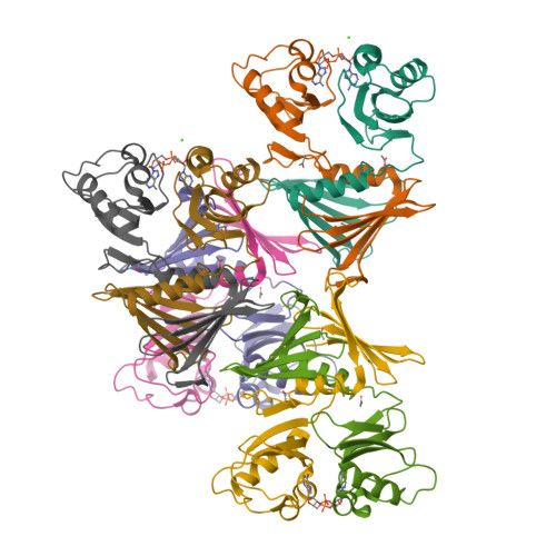
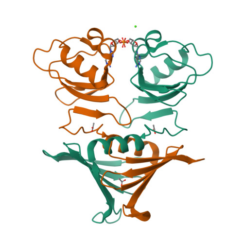
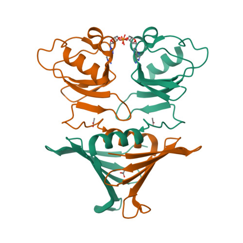
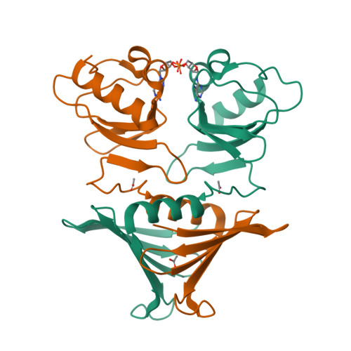
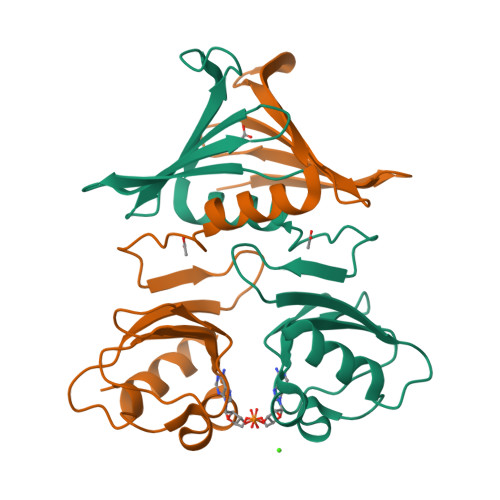
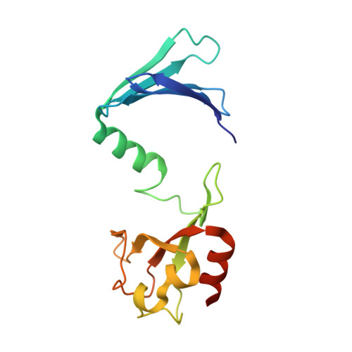
High-resolution structure of the K+/H+ antiporter subunit KhtT in complex with c-di-AMP


Experimental Data Snapshot

  • Method: X-RAY DIFFRACTION
  • Resolution: 1.85 Å
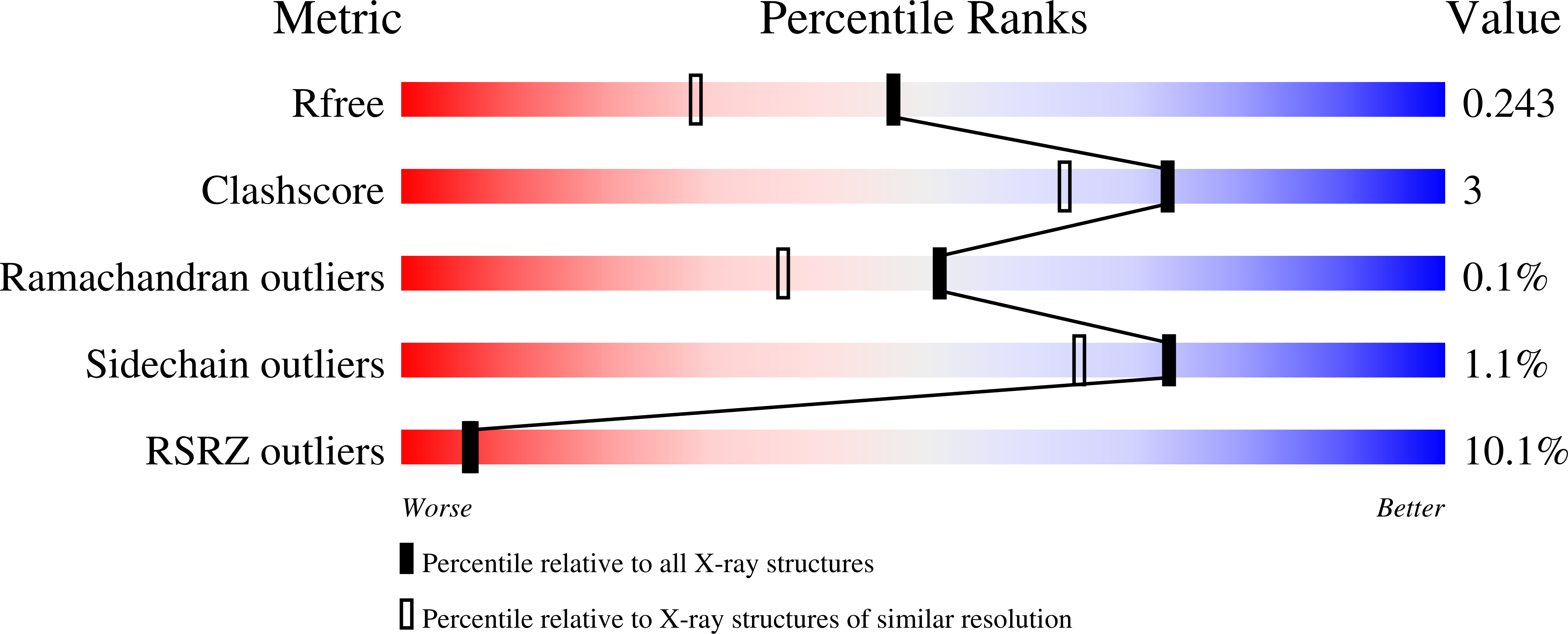
  • R-Value Free: 0.242 
  • R-Value Work: 0.204 
  • R-Value Observed: 0.206 

Starting Model: experimental
View more details

wwPDB Validation   3D Report Full Report


Ligand Structure Quality Assessment 


This is version 1.3 of the entry. See complete history


Literature

c-di-AMP, a likely master regulator of bacterial K + homeostasis machinery, activates a K + exporter.

Cereija, T.B.Guerra, J.P.L.Jorge, J.M.P.Morais-Cabral, J.H.

(2021) Proc Natl Acad Sci U S A 118

  • DOI: https://doi.org/10.1073/pnas.2020653118
  • Primary Citation of Related Structures:  
    7AGV, 7AGW, 7AGY, 7AHM, 7AHT

  • PubMed Abstract: 

    bis-(3',5')-cyclic diadenosine monophosphate (c-di-AMP) is a second messenger with roles in virulence, cell wall and biofilm formation, and surveillance of DNA integrity in many bacterial species, including pathogens. Strikingly, it has also been proposed to coordinate the activity of the components of K + homeostasis machinery, inhibiting K + import, and activating K + export. However, there is a lack of quantitative evidence supporting the direct functional impact of c-di-AMP on K + transporters. To gain a detailed understanding of the role of c-di-AMP on the activity of a component of the K + homeostasis machinery in B. subtilis , we have characterized the impact of c-di-AMP on the functional, biochemical, and physiological properties of KhtTU, a K + /H + antiporter composed of the membrane protein KhtU and the cytosolic protein KhtT. We have confirmed c-di-AMP binding to KhtT and determined the crystal structure of this complex. We have characterized in vitro the functional properties of KhtTU and KhtU alone and quantified the impact of c-di-AMP and of pH on their activity, demonstrating that c-di-AMP activates KhtTU and that pH increases its sensitivity to this nucleotide. Based on our functional and structural data, we were able to propose a mechanism for the activation of KhtTU by c-di-AMP. In addition, we have analyzed the impact of KhtTU in its native bacterium, providing a physiological context for the regulatory function of c-di-AMP and pH. Overall, we provide unique information that supports the proposal that c-di-AMP is a master regulator of K+ homeostasis machinery.


  • Organizational Affiliation

    Instituto de Biologia Molecular e Celular, Universidade do Porto, 4200-135 Porto, Portugal.


Macromolecules
Find similar proteins by:  (by identity cutoff)  |  3D Structure
Entity ID: 1
MoleculeChains Sequence LengthOrganismDetailsImage
K(+)/H(+) antiporter subunit KhtT
A, B, C, D, E
A, B, C, D, E, F, G, H
168Bacillus subtilis subsp. subtilis str. 168Mutation(s): 0 
Gene Names: khtTyhaTBSU09860
UniProt
Find proteins for O07535 (Bacillus subtilis (strain 168))
Explore O07535 
Go to UniProtKB:  O07535
Entity Groups  
Sequence Clusters30% Identity50% Identity70% Identity90% Identity95% Identity100% Identity
UniProt GroupO07535
Sequence Annotations
Expand
  • Reference Sequence
Small Molecules
Ligands 3 Unique
IDChains Name / Formula / InChI Key2D Diagram3D Interactions
2BA (Subject of Investigation/LOI)
Query on 2BA

Download Ideal Coordinates CCD File 
L [auth B],
N [auth C],
R [auth E],
X [auth H]
(2R,3R,3aS,5R,7aR,9R,10R,10aS,12R,14aR)-2,9-bis(6-amino-9H-purin-9-yl)octahydro-2H,7H-difuro[3,2-d:3',2'-j][1,3,7,9,2,8 ]tetraoxadiphosphacyclododecine-3,5,10,12-tetrol 5,12-dioxide
C20 H24 N10 O12 P2
PDXMFTWFFKBFIN-XPWFQUROSA-N
ACT
Query on ACT

Download Ideal Coordinates CCD File 
J [auth A]
K [auth A]
M [auth B]
O [auth C]
P [auth C]
J [auth A],
K [auth A],
M [auth B],
O [auth C],
P [auth C],
Q [auth D],
S [auth E],
T [auth E],
U [auth F],
W [auth G],
Y [auth H],
Z [auth H]
ACETATE ION
C2 H3 O2
QTBSBXVTEAMEQO-UHFFFAOYSA-M
CA
Query on CA

Download Ideal Coordinates CCD File 
I [auth A],
V [auth G]
CALCIUM ION
Ca
BHPQYMZQTOCNFJ-UHFFFAOYSA-N
Experimental Data & Validation

Experimental Data

  • Method: X-RAY DIFFRACTION
  • Resolution: 1.85 Å
  • R-Value Free: 0.242 
  • R-Value Work: 0.204 
  • R-Value Observed: 0.206 
  • Space Group: P 1 21 1
Unit Cell:
Length ( Å )Angle ( ˚ )
a = 72.945α = 90
b = 127.598β = 95.8
c = 81.881γ = 90
Software Package:
Software NamePurpose
PHENIXrefinement
PDB_EXTRACTdata extraction
XDSdata reduction
XDSdata scaling
PHASERphasing

Structure Validation

View Full Validation Report


Ligand Structure Quality Assessment 


Entry History & Funding Information

Deposition Data


Funding OrganizationLocationGrant Number
Fundacao para a Ciencia e a TecnologiaPortugalPOCI-01-0145-FEDER-029863(PTDC/BIA-BQM/29863/2017)

Revision History  (Full details and data files)

  • Version 1.0: 2021-03-31
    Type: Initial release
  • Version 1.1: 2021-04-07
    Changes: Database references
  • Version 1.2: 2021-04-14
    Changes: Database references
  • Version 1.3: 2024-05-01
    Changes: Data collection, Database references, Refinement description