7A7E | pdb_00007a7e

Structure of a delta-N mutant - E232start - of PA1120 (TpbB or YfiN) from Pseudomonas aeruginosa (PAO1) comprising only the GGDEF domain


Experimental Data Snapshot

  • Method: X-RAY DIFFRACTION
  • Resolution: 2.80 Å
  • R-Value Free: 
    0.224 (Depositor), 0.224 (DCC) 
  • R-Value Work: 
    0.219 (Depositor), 0.219 (DCC) 
  • R-Value Observed: 
    0.220 (Depositor) 

Starting Model: experimental
View more details

wwPDB Validation 3D Report Full Report

Validation slider image for 7A7E

This is version 1.1 of the entry. See complete history

Literature

Studying GGDEF Domain in the Act: Minimize Conformational Frustration to Prevent Artefacts.

Mantoni, F.Scribani Rossi, C.Paiardini, A.Di Matteo, A.Cappellacci, L.Petrelli, R.Ricciutelli, M.Paone, A.Cutruzzola, F.Giardina, G.Rinaldo, S.

(2021) Life (Basel) 11

  • DOI: https://doi.org/10.3390/life11010031
  • Primary Citation Related Structures: 
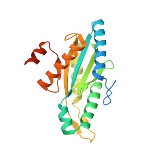
    7A7E

  • PubMed Abstract: 

    GGDEF-containing proteins respond to different environmental cues to finely modulate cyclic diguanylate (c-di-GMP) levels in time and space, making the allosteric control a distinctive trait of the corresponding proteins. The diguanylate cyclase mechanism is emblematic of this control: two GGDEF domains, each binding one GTP molecule, must dimerize to enter catalysis and yield c-di-GMP. The need for dimerization makes the GGDEF domain an ideal conformational switch in multidomain proteins. A re-evaluation of the kinetic profile of previously characterized GGDEF domains indicated that they are also able to convert GTP to GMP: this unexpected reactivity occurs when conformational issues hamper the cyclase activity. These results create new questions regarding the characterization and engineering of these proteins for in solution or structural studies.


  • Organizational Affiliation
    • Department of Biochemical Sciences "A. Rossi Fanelli", Sapienza University of Rome, Piazzale Aldo Moro, 5, 00185 Rome, Italy.

Macromolecule Content 

  • Total Structure Weight: 67.75 kDa 
  • Atom Count: 3,809 
  • Modeled Residue Count: 520 
  • Deposited Residue Count: 618 
  • Unique protein chains: 1

Macromolecules

Find similar proteins by:|  3D Structure
Entity ID: 1
MoleculeChains  Sequence LengthOrganismDetailsImage
Diguanylate cyclase TpbB
A, B, C
206Pseudomonas aeruginosa PAO1Mutation(s): 0 
Gene Names: tpbBPA1120
EC: 2.7.7.65
UniProt
Find proteins for Q9I4L5 (Pseudomonas aeruginosa (strain ATCC 15692 / DSM 22644 / CIP 104116 / JCM 14847 / LMG 12228 / 1C / PRS 101 / PAO1))
Explore Q9I4L5 
Go to UniProtKB:  Q9I4L5
Entity Groups
Sequence Clusters30% Identity50% Identity70% Identity90% Identity95% Identity100% Identity
UniProt GroupQ9I4L5
Sequence Annotations
Expand
Reference Sequence

Experimental Data & Validation

Experimental Data

  • Method: X-RAY DIFFRACTION
  • Resolution: 2.80 Å
  • R-Value Free:  0.224 (Depositor), 0.224 (DCC) 
  • R-Value Work:  0.219 (Depositor), 0.219 (DCC) 
  • R-Value Observed: 0.220 (Depositor) 
Space Group: P 21 21 21
Unit Cell:
Length ( Å )Angle ( ˚ )
a = 79.642α = 90
b = 85.104β = 90
c = 126.975γ = 90
Software Package:
Software NamePurpose
REFMACrefinement
XDSdata reduction
Aimlessdata scaling
MOLREPphasing
PDB_EXTRACTdata extraction

Structure Validation

View Full Validation Report


Entry History 

& Funding Information

Deposition Data


Funding OrganizationLocationGrant Number
Italian Ministry of EducationItalyRBFR10LHD1

Revision History  (Full details and data files)

  • Version 1.0: 2021-07-07
    Type: Initial release
  • Version 1.1: 2024-01-31
    Changes: Data collection, Database references, Refinement description