7A7B | pdb_00007a7b

Bacillithiol Disulfide Reductase Bdr (YpdA) from Staphylococcus aureus


Experimental Data Snapshot

  • Method: X-RAY DIFFRACTION
  • Resolution: 2.90 Å
  • R-Value Free: 
    0.308 (Depositor), 0.308 (DCC) 
  • R-Value Work: 
    0.243 (Depositor), 0.249 (DCC) 
  • R-Value Observed: 
    0.246 (Depositor) 

Starting Model: experimental
View more details

wwPDB Validation 3D Report Full Report

Validation slider image for 7A7B

Ligand Structure Quality Assessment 


This is version 1.3 of the entry. See complete history

Literature

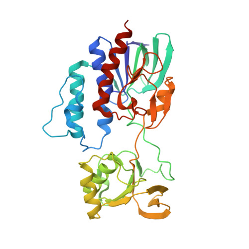
The Crystal Structures of Bacillithiol Disulfide Reductase Bdr (YpdA) Provide Structural and Functional Insight into a New Type of FAD-Containing NADPH-Dependent Oxidoreductase.

Hammerstad, M.Gudim, I.Hersleth, H.P.

(2020) Biochemistry 59: 4793-4798

  • DOI: https://doi.org/10.1021/acs.biochem.0c00745
  • Primary Citation Related Structures: 
    7A76, 7A7B, 7APR

  • PubMed Abstract: 

    Low G+C Gram-positive Firmicutes, such as the clinically important pathogens Staphylococcus aureus and Bacillus cereus , use the low-molecular weight thiol bacillithiol (BSH) as a defense mechanism to buffer the intracellular redox environment and counteract oxidative stress encountered by human neutrophils during infections. The protein YpdA has recently been shown to function as an essential NADPH-dependent reductase of oxidized bacillithiol disulfide (BSSB) resulting from stress responses and is crucial for maintaining the reduced pool of BSH and cellular redox balance. In this work, we present the first crystallographic structures of YpdAs, namely, those from S. aureus and B. cereus . Our analyses reveal a uniquely organized biological tetramer; however, the structure of the monomeric subunit is highly similar to those of other flavoprotein disulfide reductases. The absence of a redox active cysteine in the vicinity of the FAD isoalloxazine ring implies a new direct disulfide reduction mechanism, which is backed by the presence of a potentially gated channel, serving as a putative binding site for BSSB in the proximity of the FAD cofactor. We also report enzymatic activities for both YpdAs, which along with the structures presented in this work provide important structural and functional insight into a new class of FAD-containing NADPH-dependent oxidoreductases, related to the emerging fight against pathogenic bacteria.


  • Organizational Affiliation
    • Department of Biosciences, University of Oslo, Section for Biochemistry and Molecular Biology, P.O. Box 1066, Blindern, NO-0316 Oslo, Norway.

Macromolecule Content 

  • Total Structure Weight: 302.73 kDa 
  • Atom Count: 20,666 
  • Modeled Residue Count: 2,526 
  • Deposited Residue Count: 2,624 
  • Unique protein chains: 1

Macromolecules

Find similar proteins by:|  3D Structure
Entity ID: 1
MoleculeChains  Sequence LengthOrganismDetailsImage
YpdA family putative bacillithiol disulfide reductase Bdr
A, B, C, D, E
A, B, C, D, E, F, G, H
328Staphylococcus aureus subsp. aureus COLMutation(s): 0 
Gene Names: SACOL1520
UniProt
Find proteins for A0A0H2WWS2 (Staphylococcus aureus (strain COL))
Explore A0A0H2WWS2 
Go to UniProtKB:  A0A0H2WWS2
Entity Groups
Sequence Clusters30% Identity50% Identity70% Identity90% Identity95% Identity100% Identity
UniProt GroupA0A0H2WWS2
Sequence Annotations
Expand
Reference Sequence

Small Molecules

Ligands 2 Unique
IDChains Name / Formula / InChI Key2D Diagram3D Interactions
FAD
(Subject of Investigation/LOI)

Query on FAD


Download:Ideal Coordinates CCD File
I [auth A]
J [auth B]
K [auth C]
M [auth D]
O [auth E]
I [auth A],
J [auth B],
K [auth C],
M [auth D],
O [auth E],
P [auth F],
R [auth G],
S [auth H]
FLAVIN-ADENINE DINUCLEOTIDE
C27 H33 N9 O15 P2
VWWQXMAJTJZDQX-UYBVJOGSSA-N
NAP

Query on NAP


Download:Ideal Coordinates CCD File
L [auth C],
N [auth D],
Q [auth F]
NADP NICOTINAMIDE-ADENINE-DINUCLEOTIDE PHOSPHATE
C21 H28 N7 O17 P3
XJLXINKUBYWONI-NNYOXOHSSA-N

Experimental Data & Validation

Experimental Data

  • Method: X-RAY DIFFRACTION
  • Resolution: 2.90 Å
  • R-Value Free:  0.308 (Depositor), 0.308 (DCC) 
  • R-Value Work:  0.243 (Depositor), 0.249 (DCC) 
  • R-Value Observed: 0.246 (Depositor) 
Space Group: P 61 2 2
Unit Cell:
Length ( Å )Angle ( ˚ )
a = 179.315α = 90
b = 179.315β = 90
c = 349.336γ = 120
Software Package:
Software NamePurpose
PHENIXrefinement
REFMACrefinement
XDSdata reduction
Aimlessdata scaling
PHASERphasing

Structure Validation

View Full Validation Report


Ligand Structure Quality Assessment 


Entry History 

& Funding Information

Deposition Data


Funding OrganizationLocationGrant Number
Research Council of NorwayNorway231669
Research Council of NorwayNorway301584

Revision History  (Full details and data files)

  • Version 1.0: 2020-12-30
    Type: Initial release
  • Version 1.1: 2021-01-06
    Changes: Database references
  • Version 1.2: 2021-01-13
    Changes: Database references
  • Version 1.3: 2024-01-31
    Changes: Data collection, Database references, Refinement description