7A74 | pdb_00007a74

Structure of G132N BlaC from Mycobacterium tuberculosis


Experimental Data Snapshot

  • Method: X-RAY DIFFRACTION
  • Resolution: 1.60 Å
  • R-Value Free: 
    0.188 (Depositor), 0.200 (DCC) 
  • R-Value Work: 
    0.155 (Depositor), 0.170 (DCC) 
  • R-Value Observed: 
    0.157 (Depositor) 

Starting Model: experimental
View more details

wwPDB Validation 3D Report Full Report

Validation slider image for 7A74

This is version 1.4 of the entry. See complete history

Literature

Two beta-Lactamase Variants with Reduced Clavulanic Acid Inhibition Display Different Millisecond Dynamics.

Elings, W.Chikunova, A.van Zanten, D.B.Drenth, R.Ahmad, M.U.D.Blok, A.J.Timmer, M.Perrakis, A.Ubbink, M.

(2021) Antimicrob Agents Chemother 65: e0262820-e0262820

  • DOI: https://doi.org/10.1128/AAC.02628-20
  • Primary Citation Related Structures: 
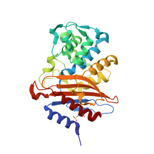
    7A74

  • PubMed Abstract: 

    The β-lactamase of Mycobacterium tuberculosis, BlaC, is susceptible to inhibition by clavulanic acid. The ability of this enzyme to escape inhibition through mutation was probed using error-prone PCR combined with functional screening in Escherichia coli. The variant that was found to confer the most inhibitor resistance, K234R, as well as variant G132N that was found previously were characterized using X-ray crystallography and nuclear magnetic resonance (NMR) relaxation experiments to probe structural and dynamic properties. The G132N mutant exists in solution in two almost equally populated conformations that exchange with a rate of ca. 88 s -1 . The conformational change affects a broad region of the enzyme. The crystal structure reveals that the Asn132 side chain forces the peptide bond between Ser104 and Ile105 in a cis -conformation. The crystal structure suggests multiple conformations for several side chains (e.g., Ser104 and Ser130) and a short loop (positions 214 to 216). In the K234R mutant, the active-site dynamics are significantly diminished with respect to the wild-type enzyme. These results show that multiple evolutionary routes are available to increase inhibitor resistance in BlaC and that active-site dynamics on the millisecond time scale are not required for catalytic function.


  • Organizational Affiliation
    • Leiden Institute of Chemistry, Leiden University, Leiden, The Netherlands.

Macromolecule Content 

  • Total Structure Weight: 29.88 kDa 
  • Atom Count: 2,368 
  • Modeled Residue Count: 267 
  • Deposited Residue Count: 267 
  • Unique protein chains: 1

Macromolecules

Find similar proteins by:|  3D Structure
Entity ID: 1
MoleculeChains  Sequence LengthOrganismDetailsImage
Beta-lactamase267Mycobacterium tuberculosis H37RvMutation(s): 1 
Gene Names: blaCblaARv2068cMTCY49.07c
EC: 3.5.2.6
UniProt
Find proteins for P9WKD3 (Mycobacterium tuberculosis (strain ATCC 25618 / H37Rv))
Explore P9WKD3 
Go to UniProtKB:  P9WKD3
Entity Groups
Sequence Clusters30% Identity50% Identity70% Identity90% Identity95% Identity100% Identity
UniProt GroupP9WKD3
Sequence Annotations
Expand
Reference Sequence

Small Molecules

Ligands 4 Unique
IDChains Name / Formula / InChI Key2D Diagram3D Interactions
PO4

Query on PO4


Download:Ideal Coordinates CCD File
R [auth A],
S [auth A]
PHOSPHATE ION
O4 P
NBIIXXVUZAFLBC-UHFFFAOYSA-K
GOL

Query on GOL


Download:Ideal Coordinates CCD File
B [auth A],
T [auth A]
GLYCEROL
C3 H8 O3
PEDCQBHIVMGVHV-UHFFFAOYSA-N
EDO

Query on EDO


Download:Ideal Coordinates CCD File
C [auth A]
D [auth A]
E [auth A]
F [auth A]
G [auth A]
C [auth A],
D [auth A],
E [auth A],
F [auth A],
G [auth A],
H [auth A],
I [auth A],
J [auth A],
K [auth A],
L [auth A],
M [auth A],
N [auth A],
O [auth A],
P [auth A],
Q [auth A],
U [auth A]
1,2-ETHANEDIOL
C2 H6 O2
LYCAIKOWRPUZTN-UHFFFAOYSA-N
NA

Query on NA


Download:Ideal Coordinates CCD File
V [auth A]SODIUM ION
Na
FKNQFGJONOIPTF-UHFFFAOYSA-N
Modified Residues  1 Unique
IDChains TypeFormula2D DiagramParent
CSD
Query on CSD
A
L-PEPTIDE LINKINGC3 H7 N O4 SCYS

Experimental Data & Validation

Experimental Data

  • Method: X-RAY DIFFRACTION
  • Resolution: 1.60 Å
  • R-Value Free:  0.188 (Depositor), 0.200 (DCC) 
  • R-Value Work:  0.155 (Depositor), 0.170 (DCC) 
  • R-Value Observed: 0.157 (Depositor) 
Space Group: P 21 21 21
Unit Cell:
Length ( Å )Angle ( ˚ )
a = 54.007α = 90
b = 54.596β = 90
c = 79.398γ = 90
Software Package:
Software NamePurpose
REFMACrefinement
XDSdata reduction
Aimlessdata scaling
MOLREPphasing

Structure Validation

View Full Validation Report


Entry History 

& Funding Information

Deposition Data


Funding OrganizationLocationGrant Number
Netherlands Organisation for Scientific Research (NWO)NetherlandsECHO-711.016.002
H2020 Marie Curie Actions of the European CommissionNetherlands653706

Revision History  (Full details and data files)

  • Version 1.0: 2021-05-19
    Type: Initial release
  • Version 1.1: 2021-06-09
    Changes: Database references, Source and taxonomy, Structure summary
  • Version 1.2: 2021-07-28
    Changes: Database references
  • Version 1.3: 2024-01-31
    Changes: Data collection, Database references, Refinement description
  • Version 1.4: 2024-10-16
    Changes: Structure summary