7A4Y

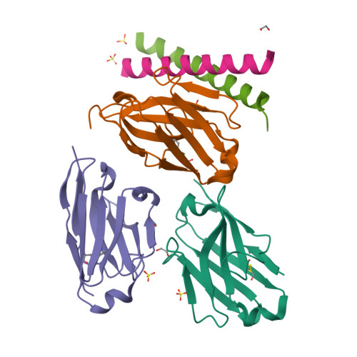
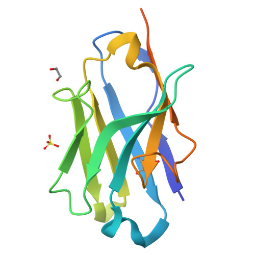
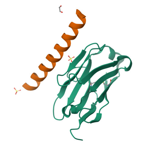
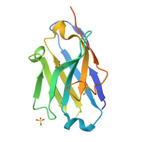
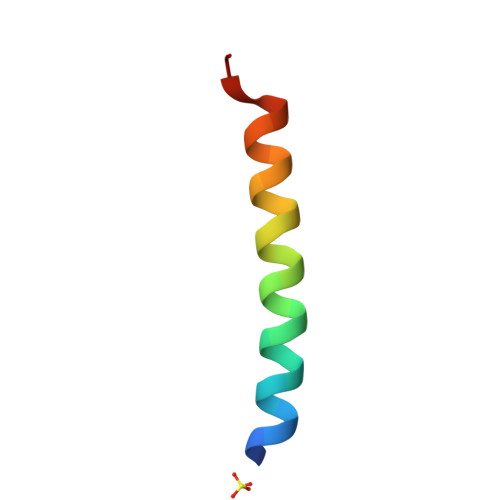
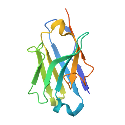
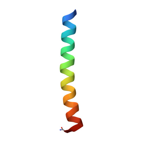
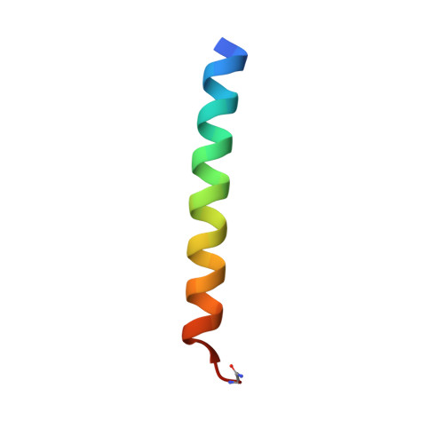
Crystal structure of the P5P6 coiled-coil in complex with nanobody Nb34.


Experimental Data Snapshot

  • Method: X-RAY DIFFRACTION
  • Resolution: 2.16 Å
  • R-Value Free: 0.229 
  • R-Value Work: 0.196 
  • R-Value Observed: 0.198 

Starting Model: in silico
View more details

wwPDB Validation   3D Report Full Report


This is version 1.2 of the entry. See complete history


Literature

A nanobody toolbox targeting dimeric coiled-coil modules for functionalization of designed protein origami structures.

Majerle, A.Hadzi, S.Aupic, J.Satler, T.Lapenta, F.Strmsek, Z.Lah, J.Loris, R.Jerala, R.

(2021) Proc Natl Acad Sci U S A 118

  • DOI: https://doi.org/10.1073/pnas.2021899118
  • Primary Citation of Related Structures:  
    7A48, 7A4D, 7A4T, 7A4Y, 7A50

  • PubMed Abstract: 

    Coiled-coil (CC) dimers are widely used in protein design because of their modularity and well-understood sequence-structure relationship. In CC protein origami design, a polypeptide chain is assembled from a defined sequence of CC building segments that determine the self-assembly of protein cages into polyhedral shapes, such as the tetrahedron, triangular prism, or four-sided pyramid. However, a targeted functionalization of the CC modules could significantly expand the versatility of protein origami scaffolds. Here, we describe a panel of single-chain camelid antibodies (nanobodies) directed against different CC modules of a de novo designed protein origami tetrahedron. We show that these nanobodies are able to recognize the same CC modules in different polyhedral contexts, such as isolated CC dimers, tetrahedra, triangular prisms, or trigonal bipyramids, thereby extending the ability to functionalize polyhedra with nanobodies in a desired stoichiometry. Crystal structures of five nanobody-CC complexes in combination with small-angle X-ray scattering show binding interactions between nanobodies and CC dimers forming the edges of a tetrahedron with the nanobody entering the tetrahedral cavity. Furthermore, we identified a pair of allosteric nanobodies in which the binding to the distant epitopes on the antiparallel homodimeric APH CC is coupled via a strong positive cooperativity. A toolbox of well-characterized nanobodies specific for CC modules provides a unique tool to target defined sites in the designed protein structures, thus opening numerous opportunities for the functionalization of CC protein origami polyhedra or CC-based bionanomaterials.


  • Organizational Affiliation

    Department of Synthetic Biology and Immunology, National Institute of Chemistry, SI-1000 Ljubljana, Slovenia.


Macromolecules
Find similar proteins by:  (by identity cutoff)  |  3D Structure
Entity ID: 1
MoleculeChains Sequence LengthOrganismDetailsImage
Nanobody Nb34
A, B, C
136Lama glamaMutation(s): 0 
Entity Groups  
Sequence Clusters30% Identity50% Identity70% Identity90% Identity95% Identity100% Identity
Sequence Annotations
Expand
  • Reference Sequence
Find similar proteins by:  (by identity cutoff)  |  3D Structure
Entity ID: 2
MoleculeChains Sequence LengthOrganismDetailsImage
Coiled-coil P6 peptide31synthetic constructMutation(s): 0 
Entity Groups  
Sequence Clusters30% Identity50% Identity70% Identity90% Identity95% Identity100% Identity
Sequence Annotations
Expand
  • Reference Sequence
Find similar proteins by:  (by identity cutoff)  |  3D Structure
Entity ID: 3
MoleculeChains Sequence LengthOrganismDetailsImage
Coiled-coil P5 peptide31synthetic constructMutation(s): 0 
Entity Groups  
Sequence Clusters30% Identity50% Identity70% Identity90% Identity95% Identity100% Identity
Sequence Annotations
Expand
  • Reference Sequence
Small Molecules
Ligands 2 Unique
IDChains Name / Formula / InChI Key2D Diagram3D Interactions
SO4
Query on SO4

Download Ideal Coordinates CCD File 
F [auth A]
H [auth B]
I [auth C]
J [auth C]
K [auth D]
F [auth A],
H [auth B],
I [auth C],
J [auth C],
K [auth D],
M [auth E]
SULFATE ION
O4 S
QAOWNCQODCNURD-UHFFFAOYSA-L
EDO
Query on EDO

Download Ideal Coordinates CCD File 
G [auth A],
L [auth D]
1,2-ETHANEDIOL
C2 H6 O2
LYCAIKOWRPUZTN-UHFFFAOYSA-N
Experimental Data & Validation

Experimental Data

  • Method: X-RAY DIFFRACTION
  • Resolution: 2.16 Å
  • R-Value Free: 0.229 
  • R-Value Work: 0.196 
  • R-Value Observed: 0.198 
  • Space Group: P 32 2 1
Unit Cell:
Length ( Å )Angle ( ˚ )
a = 72.45α = 90
b = 72.45β = 90
c = 193.8γ = 120
Software Package:
Software NamePurpose
XDSdata reduction
XSCALEdata scaling
PHASERphasing
PHENIXrefinement
PDB_EXTRACTdata extraction

Structure Validation

View Full Validation Report


Entry History & Funding Information

Deposition Data

  • Released Date: 2021-05-05 
  • Deposition Author(s): Hadzi, S.

Funding OrganizationLocationGrant Number
European Research Council (ERC)European Union787115
Slovenian Research AgencySloveniaP4-0176
Slovenian Research AgencySloveniaP1-0201

Revision History  (Full details and data files)

  • Version 1.0: 2021-05-05
    Type: Initial release
  • Version 1.1: 2024-05-01
    Changes: Data collection, Database references, Refinement description
  • Version 1.2: 2024-10-16
    Changes: Structure summary