7A1D | pdb_00007a1d

Cryo-EM map of the large glutamate dehydrogenase composed of 180 kDa subunits from Mycobacterium smegmatis (open conformation)


Experimental Data Snapshot

  • Method: ELECTRON MICROSCOPY
  • Resolution: 4.19 Å
  • Aggregation State: PARTICLE 
  • Reconstruction Method: SINGLE PARTICLE 

wwPDB Validation 3D Report Full Report

Validation slider image for 7A1D

This is version 2.0 of the entry. See complete history

Literature

3D architecture and structural flexibility revealed in the subfamily of large glutamate dehydrogenases by a mycobacterial enzyme.

Lazaro, M.Melero, R.Huet, C.Lopez-Alonso, J.P.Delgado, S.Dodu, A.Bruch, E.M.Abriata, L.A.Alzari, P.M.Valle, M.Lisa, M.N.

(2021) Commun Biol 4: 684-684

  • DOI: https://doi.org/10.1038/s42003-021-02222-x
  • Primary Citation Related Structures: 
    7A1D, 7JSR

  • PubMed Abstract: 

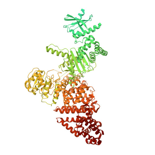
    Glutamate dehydrogenases (GDHs) are widespread metabolic enzymes that play key roles in nitrogen homeostasis. Large glutamate dehydrogenases composed of 180 kDa subunits (L-GDHs 180 ) contain long N- and C-terminal segments flanking the catalytic core. Despite the relevance of L-GDHs 180 in bacterial physiology, the lack of structural data for these enzymes has limited the progress of functional studies. Here we show that the mycobacterial L-GDH 180 (mL-GDH 180 ) adopts a quaternary structure that is radically different from that of related low molecular weight enzymes. Intersubunit contacts in mL-GDH 180 involve a C-terminal domain that we propose as a new fold and a flexible N-terminal segment comprising ACT-like and PAS-type domains that could act as metabolic sensors for allosteric regulation. These findings uncover unique aspects of the structure-function relationship in the subfamily of L-GDHs.


  • Organizational Affiliation
    • Center for Cooperative Research in Biosciences (CIC bioGUNE), Basque Research and Technology Alliance (BRTA), Bizkaia Technology Park, Building 801 A, Derio, Spain.

Macromolecule Content 

  • Total Structure Weight: 705.37 kDa 
  • Atom Count: 33,784 
  • Modeled Residue Count: 4,356 
  • Deposited Residue Count: 6,444 
  • Unique protein chains: 1

Macromolecules

Find similar proteins by:|  3D Structure
Entity ID: 1
MoleculeChains  Sequence LengthOrganismDetailsImage
NAD-specific glutamate dehydrogenase
A, B, C, D
1,611Mycolicibacterium smegmatis MC2 155Mutation(s): 0 
Gene Names: gdhMSMEG_4699MSMEI_4582
EC: 1.4.1.2
UniProt
Find proteins for A0R1C2 (Mycolicibacterium smegmatis (strain ATCC 700084 / mc(2)155))
Explore A0R1C2 
Go to UniProtKB:  A0R1C2
Entity Groups
Sequence Clusters30% Identity50% Identity70% Identity90% Identity95% Identity100% Identity
UniProt GroupA0R1C2
Sequence Annotations
Expand
Reference Sequence

Experimental Data & Validation

Experimental Data

  • Method: ELECTRON MICROSCOPY
  • Resolution: 4.19 Å
  • Aggregation State: PARTICLE 
  • Reconstruction Method: SINGLE PARTICLE 
EM Software:
TaskSoftware PackageVersion
MODEL REFINEMENTPHENIX1.21.1_5286

Structure Validation

View Full Validation Report


Entry History 

& Funding Information

Deposition Data


Funding OrganizationLocationGrant Number
Spanish Ministry of Science, Innovation, and UniversitiesSpainPGC2018-098996-B-100

Revision History  (Full details and data files)

  • Version 1.0: 2021-06-09
    Type: Initial release
  • Version 1.1: 2021-10-06
    Changes: Data collection, Database references, Structure summary
  • Version 1.2: 2024-07-10
    Changes: Data collection
  • Version 2.0: 2025-05-14
    Type: Coordinate replacement
    Reason: Model completeness
    Changes: Advisory, Atomic model, Data collection, Derived calculations, Refinement description, Source and taxonomy, Structure summary