6ZJX | pdb_00006zjx

Cold-adapted beta-D-galactosidase from Arthrobacter sp. 32cB mutant D207A in complex with saccharose


Experimental Data Snapshot

  • Method: X-RAY DIFFRACTION
  • Resolution: 2.21 Å
  • R-Value Free: 
    0.224 (Depositor), 0.225 (DCC) 
  • R-Value Work: 
    0.194 (Depositor), 0.200 (DCC) 
  • R-Value Observed: 
    0.194 (Depositor) 

Starting Model: experimental
View more details

wwPDB Validation 3D Report Full Report

Validation slider image for 6ZJX

This is version 1.2 of the entry. See complete history

Literature

Mapping the Transglycosylation Relevant Sites of Cold-Adapted beta-d-Galactosidase fromArthrobactersp. 32cB.

Rutkiewicz, M.Wanarska, M.Bujacz, A.

(2020) Int J Mol Sci 21

  • DOI: https://doi.org/10.3390/ijms21155354
  • Primary Citation Related Structures: 
    6ZJP, 6ZJQ, 6ZJR, 6ZJS, 6ZJT, 6ZJU, 6ZJV, 6ZJW, 6ZJX

  • PubMed Abstract: 

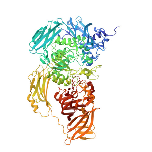
    β-Galactosidase from Arthrobacter sp. 32cB ( Arth βDG) is a cold-adapted enzyme able to catalyze hydrolysis of β-d-galactosides and transglycosylation reaction, where galactosyl moiety is being transferred onto an acceptor larger than a water molecule. Mutants of Arth βDG: D207A and E517Q were designed to determine the significance of specific residues and to enable formation of complexes with lactulose and sucrose and to shed light onto the structural basis of the transglycosylation reaction. The catalytic assays proved loss of function mutation E517 into glutamine and a significant drop of activity for mutation of D207 into alanine. Solving crystal structures of two new mutants, and new complex structures of previously presented mutant E441Q enables description of introduced changes within active site of enzyme and determining the importance of mutated residues for active site size and character. Furthermore, usage of mutants with diminished and abolished enzymatic activity enabled solving six complex structures with galactose, lactulose or sucrose bounds. As a result, not only the galactose binding sites were mapped on the enzyme's surface but also the mode of lactulose, product of transglycosylation reaction, and binding within the enzyme's active site were determined and the glucopyranose binding site in the distal of active site was discovered. The latter two especially show structural details of transglycosylation, providing valuable information that may be used for engineering of Arth βDG or other analogous galactosidases belonging to GH2 family.


  • Organizational Affiliation
    • Institute of Molecular and Industrial Biotechnology, Faculty of Biotechnology and Food Sciences, Lodz University of Technology, Stefanowskiego 4/10, 90-924 Lodz, Poland.

Macromolecule Content 

  • Total Structure Weight: 110.59 kDa 
  • Atom Count: 8,210 
  • Modeled Residue Count: 989 
  • Deposited Residue Count: 1,010 
  • Unique protein chains: 1

Macromolecules

Find similar proteins by:|  3D Structure
Entity ID: 1
MoleculeChains  Sequence LengthOrganismDetailsImage
Beta-galactosidase1,010Arthrobacter sp. 32cBMutation(s): 1 
EC: 3.2.1.23
UniProt
Find proteins for A0A023UGN9 (Arthrobacter sp. 32cB)
Explore A0A023UGN9 
Go to UniProtKB:  A0A023UGN9
Entity Groups
Sequence Clusters30% Identity50% Identity70% Identity90% Identity95% Identity100% Identity
UniProt GroupA0A023UGN9
Sequence Annotations
Expand
Reference Sequence

Oligosaccharides

Help  
Entity ID: 2
MoleculeChains Length2D Diagram GlycosylationD Interactions
beta-D-fructofuranose-(2-1)-alpha-D-glucopyranose
B, C
2N/A
Glycosylation Resources
GlyTouCan: G05551OP
GlyCosmos: G05551OP

Biologically Interesting Molecules (External Reference) 

1 Unique

Experimental Data & Validation

Experimental Data

  • Method: X-RAY DIFFRACTION
  • Resolution: 2.21 Å
  • R-Value Free:  0.224 (Depositor), 0.225 (DCC) 
  • R-Value Work:  0.194 (Depositor), 0.200 (DCC) 
  • R-Value Observed: 0.194 (Depositor) 
Space Group: P 31 2 1
Unit Cell:
Length ( Å )Angle ( ˚ )
a = 137.574α = 90
b = 137.574β = 90
c = 126.81γ = 120
Software Package:
Software NamePurpose
PHENIXrefinement
XSCALEdata scaling
PDB_EXTRACTdata extraction
XDSdata reduction
PHENIXphasing

Structure Validation

View Full Validation Report


Entry History 

& Funding Information

Deposition Data


Funding OrganizationLocationGrant Number
Polish National Science CentrePoland2016/21/B/ST5/00555
Polish National Science CentrePoland2018/28/T/ST5/00233

Revision History  (Full details and data files)

  • Version 1.0: 2020-08-05
    Type: Initial release
  • Version 1.1: 2020-08-12
    Changes: Database references
  • Version 1.2: 2024-01-31
    Changes: Data collection, Database references, Refinement description