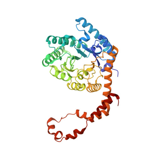
Attaining atomic resolution from in situ data collection at room temperature using counter-diffusion-based low-cost microchips.
Gavira, J.A., Rodriguez-Ruiz, I., Martinez-Rodriguez, S., Basu, S., Teychene, S., McCarthy, A.A., Mueller-Dieckman, C.(2020) Acta Crystallogr D Struct Biol 76: 751-758
- PubMed: 32744257
- DOI: https://doi.org/10.1107/S2059798320008475
- Primary Citation of Related Structures:
6YBF, 6YBI, 6YBO, 6YBR, 6YBX, 6YC5 - PubMed Abstract:
Sample handling and manipulation for cryoprotection currently remain critical factors in X-ray structural determination. While several microchips for macromolecular crystallization have been proposed during the last two decades to partially overcome crystal-manipulation issues, increased background noise originating from the scattering of chip-fabrication materials has so far limited the attainable resolution of diffraction data. Here, the conception and use of low-cost, X-ray-transparent microchips for in situ crystallization and direct data collection, and structure determination at atomic resolution close to 1.0 Å, is presented. The chips are fabricated by a combination of either OSTEMER and Kapton or OSTEMER and Mylar materials for the implementation of counter-diffusion crystallization experiments. Both materials produce a sufficiently low scattering background to permit atomic resolution diffraction data collection at room temperature and the generation of 3D structural models of the tested model proteins lysozyme, thaumatin and glucose isomerase. Although the high symmetry of the three model protein crystals produced almost complete data sets at high resolution, the potential of in-line data merging and scaling of the multiple crystals grown along the microfluidic channels is also presented and discussed.
- Laboratorio de Estudios Cristalográficos, IACT, CSIC-Universidad de Granada, Avenida Las Palmeras 4, 18100 Armilla, Spain.
Organizational Affiliation:


