6Y32 | pdb_00006y32

Structure of the GTPase heterodimer of human SRP54 and SRalpha


Experimental Data Snapshot

  • Method: X-RAY DIFFRACTION
  • Resolution: 2.60 Å
  • R-Value Free: 
    0.184 (Depositor), 0.173 (DCC) 
  • R-Value Work: 
    0.149 (Depositor), 0.151 (DCC) 
  • R-Value Observed: 
    0.154 (Depositor) 

Starting Model: experimental
View more details

wwPDB Validation 3D Report Full Report

Validation slider image for 6Y32

Ligand Structure Quality Assessment 


This is version 1.4 of the entry. See complete history

Literature

Structural and Functional Impact of SRP54 Mutations Causing Severe Congenital Neutropenia.

Juaire, K.D.Lapouge, K.Becker, M.M.M.Kotova, I.Michelhans, M.Carapito, R.Wild, K.Bahram, S.Sinning, I.

(2021) Structure 29: 15

  • DOI: https://doi.org/10.1016/j.str.2020.09.008
  • Primary Citation Related Structures: 
    6Y2Z, 6Y30, 6Y31, 6Y32

  • PubMed Abstract: 

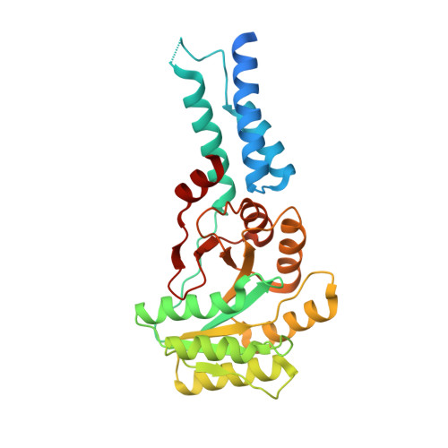
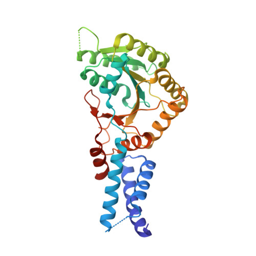
    The SRP54 GTPase is a key component of co-translational protein targeting by the signal recognition particle (SRP). Point mutations in SRP54 have been recently shown to lead to a form of severe congenital neutropenia displaying symptoms overlapping with those of Shwachman-Diamond syndrome. The phenotype includes severe neutropenia, exocrine pancreatic deficiency, and neurodevelopmental as well as skeletal disorders. Using a combination of X-ray crystallography, hydrogen-deuterium exchange coupled to mass spectrometry and complementary biochemical and biophysical methods, we reveal extensive structural defects in three disease-causing SRP54 variants resulting in critical protein destabilization. GTP binding is mostly abolished as a consequence of an altered GTPase core. The mutations located in conserved sequence fingerprints of SRP54 eliminate targeting complex formation with the SRP receptor as demonstrated in yeast and human cells. These specific defects critically influence the entire SRP pathway, thereby causing this life-threatening disease.


  • Organizational Affiliation
    • Heidelberg University Biochemistry Center (BZH), Im Neuenheimer Feld 328, D-69120 Heidelberg, Germany.

Macromolecule Content 

  • Total Structure Weight: 277.7 kDa 
  • Atom Count: 18,356 
  • Modeled Residue Count: 2,288 
  • Deposited Residue Count: 2,476 
  • Unique protein chains: 2

Macromolecules

Find similar proteins by:|  3D Structure
Entity ID: 1
MoleculeChains  Sequence LengthOrganismDetailsImage
Signal recognition particle 54 kDa protein
A, C, E, G
304Homo sapiensMutation(s): 0 
Gene Names: SRP54
EC: 3.6.5.4
UniProt & NIH Common Fund Data Resources
Find proteins for P61011 (Homo sapiens)
Explore P61011 
Go to UniProtKB:  P61011
PHAROS:  P61011
GTEx:  ENSG00000100883 
Entity Groups
Sequence Clusters30% Identity50% Identity70% Identity90% Identity95% Identity100% Identity
UniProt GroupP61011
Sequence Annotations
Expand
Reference Sequence
Find similar proteins by:|  3D Structure
Entity ID: 2
MoleculeChains  Sequence LengthOrganismDetailsImage
Signal recognition particle receptor subunit alpha
B, D, F, H
315Homo sapiensMutation(s): 0 
Gene Names: SRPRASRPR
UniProt & NIH Common Fund Data Resources
Find proteins for P08240 (Homo sapiens)
Explore P08240 
Go to UniProtKB:  P08240
PHAROS:  P08240
GTEx:  ENSG00000182934 
Entity Groups
Sequence Clusters30% Identity50% Identity70% Identity90% Identity95% Identity100% Identity
UniProt GroupP08240
Sequence Annotations
Expand
Reference Sequence

Small Molecules

Ligands 3 Unique
IDChains Name / Formula / InChI Key2D Diagram3D Interactions
GNP
(Subject of Investigation/LOI)

Query on GNP


Download:Ideal Coordinates CCD File
CA [auth G]
FA [auth H]
I [auth A]
K [auth B]
P [auth C]
CA [auth G],
FA [auth H],
I [auth A],
K [auth B],
P [auth C],
S [auth D],
V [auth E],
X [auth F]
PHOSPHOAMINOPHOSPHONIC ACID-GUANYLATE ESTER
C10 H17 N6 O13 P3
UQABYHGXWYXDTK-UUOKFMHZSA-N
SO4

Query on SO4


Download:Ideal Coordinates CCD File
AA [auth F]
BA [auth F]
EA [auth G]
HA [auth H]
IA [auth H]
AA [auth F],
BA [auth F],
EA [auth G],
HA [auth H],
IA [auth H],
JA [auth H],
KA [auth H],
LA [auth H],
M [auth B],
N [auth B],
O [auth B],
R [auth C],
U [auth D],
Z [auth F]
SULFATE ION
O4 S
QAOWNCQODCNURD-UHFFFAOYSA-L
MG

Query on MG


Download:Ideal Coordinates CCD File
DA [auth G]
GA [auth H]
J [auth A]
L [auth B]
Q [auth C]
DA [auth G],
GA [auth H],
J [auth A],
L [auth B],
Q [auth C],
T [auth D],
W [auth E],
Y [auth F]
MAGNESIUM ION
Mg
JLVVSXFLKOJNIY-UHFFFAOYSA-N

Experimental Data & Validation

Experimental Data

  • Method: X-RAY DIFFRACTION
  • Resolution: 2.60 Å
  • R-Value Free:  0.184 (Depositor), 0.173 (DCC) 
  • R-Value Work:  0.149 (Depositor), 0.151 (DCC) 
  • R-Value Observed: 0.154 (Depositor) 
Space Group: P 1 2 1
Unit Cell:
Length ( Å )Angle ( ˚ )
a = 155.825α = 90
b = 70.634β = 116.81
c = 172.573γ = 90
Software Package:
Software NamePurpose
PHENIXrefinement
PDB_EXTRACTdata extraction
XDSdata reduction
Aimlessdata scaling
PHENIXphasing

Structure Validation

View Full Validation Report


Ligand Structure Quality Assessment 


Entry History 

& Funding Information

Deposition Data


Funding OrganizationLocationGrant Number
German Research Foundation (DFG)GermanySI 586/6-1

Revision History  (Full details and data files)

  • Version 1.0: 2020-09-23
    Type: Initial release
  • Version 1.1: 2020-12-16
    Changes: Database references
  • Version 1.2: 2021-01-20
    Changes: Database references
  • Version 1.3: 2024-01-24
    Changes: Data collection, Database references, Refinement description
  • Version 1.4: 2024-10-16
    Changes: Structure summary