6X5I

Cryo-EM of peptide-like filament of 1-KMe3


Experimental Data Snapshot

  • Method: ELECTRON MICROSCOPY
  • Resolution: 4.30 Å
  • Aggregation State: FILAMENT 
  • Reconstruction Method: HELICAL 

wwPDB Validation   3D Report Full Report

Currently 6X5I does not have a validation slider image.


This is version 2.1 of the entry. See complete history


Literature

Artificial Intracellular Filaments.

Feng, Z.Wang, H.Wang, F.Oh, Y.Berciu, C.Cui, Q.Egelman, E.H.Xu, B.

(2020) Cell Rep Phys Sci 1

  • DOI: https://doi.org/10.1016/j.xcrp.2020.100085
  • Primary Citation of Related Structures:  
    6X5I

  • PubMed Abstract: 

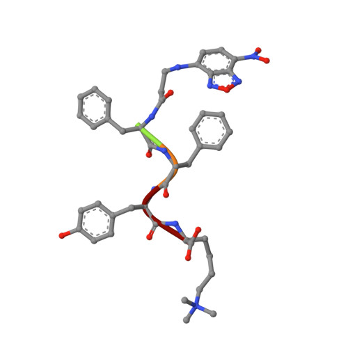
    Intracellular protein filaments are ubiquitous for cellular functions, but forming bona fide biomimetic intracellular filaments of small molecules in living cells remains elusive. Here, we report the in situ formation of self-limiting intracellular filaments of a small peptide via enzymatic morphological transition of a phosphorylated and trimethylated heterochiral tetrapeptide. Enzymatic dephosphorylation reduces repulsive intermolecular electrostatic interactions and converts the peptidic nanoparticles into filaments, which exhibit distinct types of cross-β structures with either C7 or C2 symmetries, with the hydrophilic C-terminal residues at the periphery of the helix. Macromolecular crowding promotes the peptide filaments to form bundles, which extend from the plasma membrane to nuclear membrane and hardly interact with endogenous components, including cytoskeletons. Stereochemistry and post-translational modification (PTM) of peptides are critical for generating the intracellular bundles. This work may offer a way to gain lost functions or to provide molecular insights for understanding normal and aberrant intracellular filaments.


  • Organizational Affiliation

    Department of Chemistry, Brandeis University, 415 South Street, Waltham, MA 02454, USA.


Macromolecules

Find similar proteins by:  Sequence   |   3D Structure  

Entity ID: 1
MoleculeChains Sequence LengthOrganismDetailsImage
1-KMe3 peptide-like fibril5Homo sapiensMutation(s): 0 
Sequence Annotations
Expand
  • Reference Sequence
Small Molecules
Modified Residues  1 Unique
IDChains TypeFormula2D DiagramParent
M3L
Query on M3L
A [auth B]
AA [auth c]
AB [auth BA]
B [auth D]
BA [auth d]
A [auth B],
AA [auth c],
AB [auth BA],
B [auth D],
BA [auth d],
BB [auth CA],
C [auth E],
CA [auth e],
CB [auth DA],
D [auth F],
DA [auth f],
DB [auth EA],
E [auth G],
EA [auth g],
EB [auth FA],
F [auth H],
FA [auth h],
FB [auth GA],
G [auth I],
GA [auth i],
GB [auth HA],
H [auth J],
HA [auth j],
HB [auth IA],
I [auth K],
IA [auth k],
IB [auth JA],
J [auth L],
JA [auth l],
JB [auth KA],
K [auth M],
KA [auth m],
KB [auth LA],
L [auth N],
LA [auth n],
LB [auth MA],
M [auth O],
MA [auth o],
MB [auth NA],
N [auth P],
NA [auth p],
NB [auth OA],
O [auth Q],
OA [auth q],
OB [auth PA],
P [auth R],
PA [auth r],
PB [auth QA],
Q [auth S],
QA [auth s],
QB [auth RA],
R [auth T],
RA [auth t],
RB [auth SA],
S [auth U],
SA [auth u],
T [auth V],
TA [auth v],
U [auth W],
UA [auth w],
V [auth X],
VA [auth x],
W [auth Y],
WA [auth y],
X [auth Z],
XA [auth z],
Y [auth a],
YA [auth 0],
Z [auth b],
ZA [auth AA]
L-PEPTIDE LINKINGC9 H21 N2 O2LYS
Experimental Data & Validation

Experimental Data

  • Method: ELECTRON MICROSCOPY
  • Resolution: 4.30 Å
  • Aggregation State: FILAMENT 
  • Reconstruction Method: HELICAL 

Structure Validation

View Full Validation Report

Currently 6X5I does not have a validation slider image.


Entry History & Funding Information

Deposition Data


Funding OrganizationLocationGrant Number
National Institutes of Health/National Institute of General Medical Sciences (NIH/NIGMS)United StatesR35GM122510

Revision History  (Full details and data files)

  • Version 1.0: 2020-06-03
    Type: Initial release
  • Version 2.0: 2020-07-15
    Changes: Database references, Non-polymer description
  • Version 2.1: 2020-08-26
    Changes: Database references