6V4B | pdb_00006v4b

DeCLIC N-terminal Domain 34-202


Experimental Data Snapshot

  • Method: X-RAY DIFFRACTION
  • Resolution: 1.75 Å
  • R-Value Free: 
    0.220 (Depositor), 0.229 (DCC) 
  • R-Value Work: 
    0.199 (Depositor), 0.203 (DCC) 
  • R-Value Observed: 
    0.200 (Depositor) 

wwPDB Validation 3D Report Full Report

Validation slider image for 6V4B

This is version 1.3 of the entry. See complete history

Literature

Structural basis for allosteric transitions of a multidomain pentameric ligand-gated ion channel.

Hu, H.Howard, R.J.Bastolla, U.Lindahl, E.Delarue, M.

(2020) Proc Natl Acad Sci U S A 117: 13437-13446

  • DOI: https://doi.org/10.1073/pnas.1922701117
  • Primary Citation Related Structures: 
    6V4A, 6V4B, 6V4S

  • PubMed Abstract: 

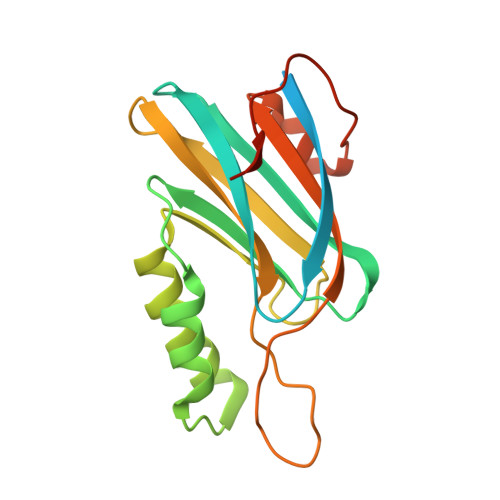
    Pentameric ligand-gated ion channels (pLGICs) are allosteric receptors that mediate rapid electrochemical signal transduction in the animal nervous system through the opening of an ion pore upon binding of neurotransmitters. Orthologs have been found and characterized in prokaryotes and they display highly similar structure-function relationships to eukaryotic pLGICs; however, they often encode greater architectural diversity involving additional amino-terminal domains (NTDs). Here we report structural, functional, and normal-mode analysis of two conformational states of a multidomain pLGIC, called DeCLIC, from a Desulfofustis deltaproteobacterium, including a periplasmic NTD fused to the conventional ligand-binding domain (LBD). X-ray structure determination revealed an NTD consisting of two jelly-roll domains interacting across each subunit interface. Binding of Ca 2+ at the LBD subunit interface was associated with a closed transmembrane pore, with resolved monovalent cations intracellular to the hydrophobic gate. Accordingly, DeCLIC-injected oocytes conducted currents only upon depletion of extracellular Ca 2+ ; these were insensitive to quaternary ammonium block. Furthermore, DeCLIC crystallized in the absence of Ca 2+ with a wide-open pore and remodeled periplasmic domains, including increased contacts between the NTD and classic LBD agonist-binding sites. Functional, structural, and dynamical properties of DeCLIC paralleled those of sTeLIC, a pLGIC from another symbiotic prokaryote. Based on these DeCLIC structures, we would reclassify the previous structure of bacterial ELIC (the first high-resolution structure of a pLGIC) as a "locally closed" conformation. Taken together, structures of DeCLIC in multiple conformations illustrate dramatic conformational state transitions and diverse regulatory mechanisms available to ion channels in pLGICs, particularly involving Ca 2+ modulation and periplasmic NTDs.


  • Organizational Affiliation
    • Unité Dynamique Structurale des Macromolécules, Institut Pasteur, UMR 3528, CNRS, 75015 Paris, France.

Macromolecule Content 

  • Total Structure Weight: 45.44 kDa 
  • Atom Count: 2,833 
  • Modeled Residue Count: 320 
  • Deposited Residue Count: 404 
  • Unique protein chains: 1

Macromolecules

Find similar proteins by:|  3D Structure
Entity ID: 1
MoleculeChains  Sequence LengthOrganismDetailsImage
Neur_chan_LBD domain-containing protein
A, B
202Desulfofustis sp. PB-SRB1Mutation(s): 0 
Gene Names: N839_03575
UniProt
Find proteins for A0ACD6B9Z9 (Desulfofustis sp. PB-SRB1)
Explore A0ACD6B9Z9 
Go to UniProtKB:  A0ACD6B9Z9
Entity Groups
Sequence Clusters30% Identity50% Identity70% Identity90% Identity95% Identity100% Identity
UniProt GroupA0ACD6B9Z9
Sequence Annotations
Expand
Reference Sequence

Small Molecules

Ligands 3 Unique
IDChains Name / Formula / InChI Key2D Diagram3D Interactions
EPE

Query on EPE


Download:Ideal Coordinates CCD File
E [auth A],
J [auth B]
4-(2-HYDROXYETHYL)-1-PIPERAZINE ETHANESULFONIC ACID
C8 H18 N2 O4 S
JKMHFZQWWAIEOD-UHFFFAOYSA-N
PO4

Query on PO4


Download:Ideal Coordinates CCD File
F [auth A],
G [auth A],
K [auth B],
L [auth B]
PHOSPHATE ION
O4 P
NBIIXXVUZAFLBC-UHFFFAOYSA-K
GOL

Query on GOL


Download:Ideal Coordinates CCD File
C [auth A],
D [auth A],
H [auth B],
I [auth B]
GLYCEROL
C3 H8 O3
PEDCQBHIVMGVHV-UHFFFAOYSA-N

Experimental Data & Validation

Experimental Data

  • Method: X-RAY DIFFRACTION
  • Resolution: 1.75 Å
  • R-Value Free:  0.220 (Depositor), 0.229 (DCC) 
  • R-Value Work:  0.199 (Depositor), 0.203 (DCC) 
  • R-Value Observed: 0.200 (Depositor) 
Space Group: P 61
Unit Cell:
Length ( Å )Angle ( ˚ )
a = 81.35α = 90
b = 81.35β = 90
c = 99.26γ = 120
Software Package:
Software NamePurpose
BUSTERrefinement
XDSdata reduction
Aimlessdata scaling
PHASERphasing

Structure Validation

View Full Validation Report


Entry History 

Deposition Data

Revision History  (Full details and data files)

  • Version 1.0: 2020-06-03
    Type: Initial release
  • Version 1.1: 2020-06-17
    Changes: Database references
  • Version 1.2: 2020-07-01
    Changes: Database references
  • Version 1.3: 2024-03-06
    Changes: Data collection, Database references