6UWM

Single particle cryo-EM structure of KvAP


Experimental Data Snapshot

  • Method: ELECTRON MICROSCOPY
  • Resolution: 5.90 Å
  • Aggregation State: PARTICLE 
  • Reconstruction Method: SINGLE PARTICLE 

wwPDB Validation   3D Report Full Report


This is version 1.2 of the entry. See complete history


Literature

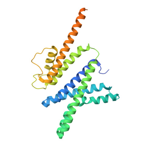
Cryo-EM structure of the KvAP channel reveals a non-domain-swapped voltage sensor topology.

Tao, X.MacKinnon, R.

(2019) Elife 8

  • DOI: https://doi.org/10.7554/eLife.52164
  • Primary Citation of Related Structures:  
    6UWM

  • PubMed Abstract: 

    Conductance in voltage-gated ion channels is regulated by membrane voltage through structural domains known as voltage sensors. A single structural class of voltage sensor domain exists, but two different modes of voltage sensor attachment to the pore occur in nature: domain-swapped and non-domain-swapped. Since the more thoroughly studied Kv1-7, Nav and Cav channels have domain-swapped voltage sensors, much less is known about non-domain-swapped voltage-gated ion channels. In this paper, using cryo-EM, we show that KvAP from Aeropyrum pernix has non-domain-swapped voltage sensors as well as other unusual features. The new structure, together with previous functional data, suggests that KvAP and the Shaker channel, to which KvAP is most often compared, probably undergo rather different voltage-dependent conformational changes when they open.


  • Organizational Affiliation

    Laboratory of Molecular Neurobiology and Biophysics, The Rockefeller University, Howard Hughes Medical Institute, New York, United States.


Macromolecules
Find similar proteins by:  (by identity cutoff)  |  3D Structure
Entity ID: 1
MoleculeChains Sequence LengthOrganismDetailsImage
Voltage-gated potassium channelA,
B [auth C],
C [auth B],
D
287Aeropyrum pernixMutation(s): 0 
Gene Names: APE_0955
Membrane Entity: Yes 
UniProt
Find proteins for Q9YDF8 (Aeropyrum pernix (strain ATCC 700893 / DSM 11879 / JCM 9820 / NBRC 100138 / K1))
Explore Q9YDF8 
Go to UniProtKB:  Q9YDF8
Entity Groups  
Sequence Clusters30% Identity50% Identity70% Identity90% Identity95% Identity100% Identity
UniProt GroupQ9YDF8
Sequence Annotations
Expand
  • Reference Sequence
Experimental Data & Validation

Experimental Data

  • Method: ELECTRON MICROSCOPY
  • Resolution: 5.90 Å
  • Aggregation State: PARTICLE 
  • Reconstruction Method: SINGLE PARTICLE 
EM Software:
TaskSoftware PackageVersion
MODEL REFINEMENTPHENIX1.16
RECONSTRUCTIONFREALIGN9.11

Structure Validation

View Full Validation Report


Entry History & Funding Information

Deposition Data


Funding OrganizationLocationGrant Number
National Institutes of Health/National Institute of General Medical Sciences (NIH/NIGMS)United StatesGM43949

Revision History  (Full details and data files)

  • Version 1.0: 2019-12-04
    Type: Initial release
  • Version 1.1: 2020-01-08
    Changes: Author supporting evidence
  • Version 1.2: 2024-03-06
    Changes: Data collection, Database references, Refinement description