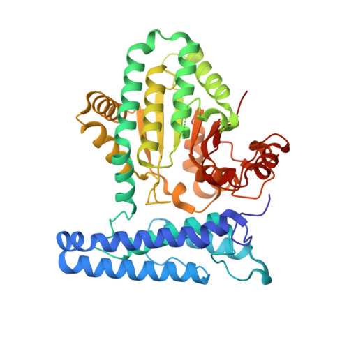
6UMQ

Structure of DUF89


Experimental Data Snapshot

  • Method: X-RAY DIFFRACTION
  • Resolution: 1.85 Å
  • R-Value Free: 0.224 
  • R-Value Work: 0.196 
  • R-Value Observed: 0.197 

Starting Model: experimental
View more details

wwPDB Validation   3D Report Full Report

Currently 6UMQ does not have a validation slider image.


This is version 1.2 of the entry. See complete history


Literature

Human ARMT1 structure and substrate specificity indicates that it is a DUF89 family damage-control phosphatase.

Dennis, T.N.Kenjic, N.Kang, A.S.Lowenson, J.D.Kirkwood, J.S.Clarke, S.G.Perry, J.J.P.

(2020) J Struct Biol 212: 107576-107576

  • DOI: https://doi.org/10.1016/j.jsb.2020.107576
  • Primary Citation of Related Structures:  
    6UMQ, 6UMR

  • PubMed Abstract: 

    Metabolite damage control is a critical but poorly defined aspect of cellular biochemistry, which likely involves many of the so far functionally uncharacterized protein domain (domains of unknown function; DUFs). We have determined the crystal structure of the human DUF89 protein product of the C6ORF211 gene to 1.85 Å. The crystal structure shows that the protein contains a core α-β-α fold with an active site-bound metal ion and α-helical bundle N-terminal cap, which are both conserved features of subfamily III DUF89 domains. The biochemical activities of the human protein are conserved with those of a previously characterized budding yeast homolog, where an in vitro phosphatase activity is supported by divalent cations that include Co 2+ , Ni 2+ , Mn 2+ or Mg 2+ . Full steady-state kinetics parameters of human DUF89 using a standard PNPP phosphatase assay revealed a six times higher catalytic efficiency in presence of Co 2+ compared to Mg 2+ . The human enzyme targets a number of phosphate substrates similar to the budding yeast homolog, while it lacks a previously indicated methyltransferase activity. The highest activity on substrate was observed with fructose-1-phosphate, a potent glycating agent, and thus human DUF89 phosphatase activity may also play a role in limiting the buildup of phospho-glycan species and their related damaged metabolites.


  • Organizational Affiliation

    Department of Biochemistry, University of California, Riverside, Riverside, CA 92521, USA.


Macromolecules
Find similar proteins by:  (by identity cutoff)  |  3D Structure
Entity ID: 1
MoleculeChains Sequence LengthOrganismDetailsImage
Damage-control phosphatase DUF89
A, B
441Homo sapiensMutation(s): 0 
Gene Names: ARMT1C6orf211
EC: 3.1.3 (PDB Primary Data), 2.1.1 (PDB Primary Data)
UniProt & NIH Common Fund Data Resources
Find proteins for Q9H993 (Homo sapiens)
Explore Q9H993 
Go to UniProtKB:  Q9H993
PHAROS:  Q9H993
GTEx:  ENSG00000146476 
Entity Groups  
Sequence Clusters30% Identity50% Identity70% Identity90% Identity95% Identity100% Identity
UniProt GroupQ9H993
Sequence Annotations
Expand
  • Reference Sequence
Experimental Data & Validation

Experimental Data

  • Method: X-RAY DIFFRACTION
  • Resolution: 1.85 Å
  • R-Value Free: 0.224 
  • R-Value Work: 0.196 
  • R-Value Observed: 0.197 
  • Space Group: C 2 2 21
Unit Cell:
Length ( Å )Angle ( ˚ )
a = 90.14α = 90
b = 194.473β = 90
c = 114.201γ = 90
Software Package:
Software NamePurpose
PHENIXrefinement
MOSFLMdata reduction
SCALAdata scaling
Cootmodel building
PHENIXphasing

Structure Validation

View Full Validation Report

Currently 6UMQ does not have a validation slider image.


Entry History & Funding Information

Deposition Data


Funding OrganizationLocationGrant Number
National Science Foundation (NSF, United States)United StatesMCB-1714569

Revision History  (Full details and data files)

  • Version 1.0: 2020-07-29
    Type: Initial release
  • Version 1.1: 2020-10-07
    Changes: Database references
  • Version 1.2: 2023-10-11
    Changes: Data collection, Database references, Refinement description