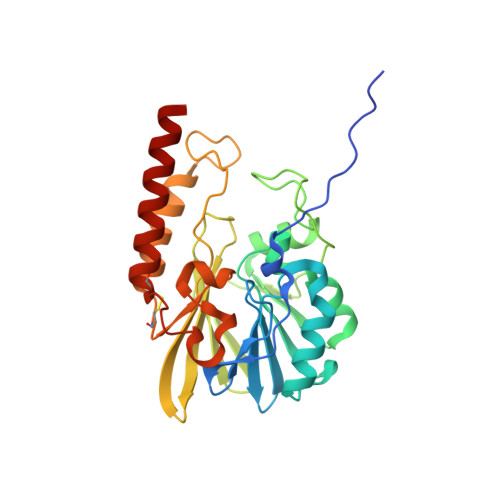
Structural and biochemical analysis of the metallo-beta-lactamase L1 from emerging pathogen Stenotrophomonas maltophilia revealed the subtle but distinct di-metal scaffold for catalytic activity.
Kim, Y., Maltseva, N., Wilamowski, M., Tesar, C., Endres, M., Joachimiak, A.(2020) Protein Sci 29: 723-743
- PubMed: 31846104 Search on PubMedSearch on PubMed Central
- DOI: https://doi.org/10.1002/pro.3804
- Primary Citation Related Structures:
6U0Y, 6U0Z, 6U10, 6U13, 6U2Z, 6U9C, 6UA1, 6UAC, 6UAF, 6UAH - PubMed Abstract:
Emergence of Enterobacteriaceae harboring metallo-β-lactamases (MBL) has raised global threats due to their broad antibiotic resistance profiles and the lack of effective inhibitors against them. We have been studied one of the emerging environmental MBL, the L1 from Stenotrophomonas maltophilia K279a. We determined several crystal structures of L1 complexes with three different classes of β-lactam antibiotics (penicillin G, moxalactam, meropenem, and imipenem), with the inhibitor captopril and different metal ions (Zn +2 , Cd +2 , and Cu +2 ). All hydrolyzed antibiotics and the inhibitor were found binding to two Zn +2 ions mainly through the opened lactam ring and some hydrophobic interactions with the binding pocket atoms. Without a metal ion, the active site is very similarly maintained as that of the native form with two Zn +2 ions, however, the protein does not bind the substrate moxalactam. When two Zn +2 ions were replaced with other metal ions, the same di-metal scaffold was maintained and the added moxalactam was found hydrolyzed in the active site. Differential scanning fluorimetry and isothermal titration calorimetry were used to study thermodynamic properties of L1 MBL compared with New Deli Metallo-β-lactamase-1 (NDM-1). Both enzymes are significantly stabilized by Zn +2 and other divalent metals but showed different dependency. These studies also suggest that moxalactam and its hydrolyzed form may bind and dissociate with different kinetic modes with or without Zn +2 for each of L1 and NDM-1. Our analysis implicates metal ions, in forming a distinct di-metal scaffold, which is central to the enzyme stability, promiscuous substrate binding and versatile catalytic activity. STATEMENT: The L1 metallo-β-lactamase from an environmental multidrug-resistant opportunistic pathogen Stenotrophomonas maltophilia K279a has been studied by determining 3D structures of L1 enzyme in the complexes with several β-lactam antibiotics and different divalent metals and characterizing its biochemical and ligand binding properties. We found that the two-metal center in the active site is critical in the enzymatic process including antibiotics recognition and binding, which explains the enzyme's activity toward diverse antibiotic substrates. This study provides the critical information for understanding the ligand recognition and for advanced drug development.
- Center for Structural Genomics of Infectious Diseases, Consortium for Advanced Science and Engineering, the University of Chicago, Chicago, Illinois.
Organizational Affiliation: