6RAP

Cryo-EM structure of the anti-feeding prophage cap (AFP tube terminating cap)


Experimental Data Snapshot

  • Method: ELECTRON MICROSCOPY
  • Resolution: 3.30 Å
  • Aggregation State: PARTICLE 
  • Reconstruction Method: SINGLE PARTICLE 

wwPDB Validation   3D Report Full Report


This is version 1.2 of the entry. See complete history


Literature

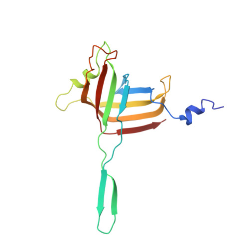
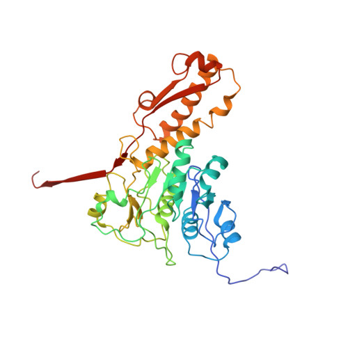
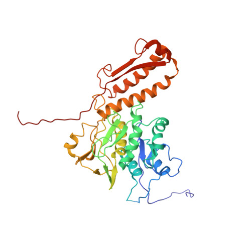
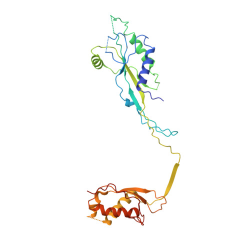
Atomic structures of an entire contractile injection system in both the extended and contracted states.

Desfosses, A.Venugopal, H.Joshi, T.Felix, J.Jessop, M.Jeong, H.Hyun, J.Heymann, J.B.Hurst, M.R.H.Gutsche, I.Mitra, A.K.

(2019) Nat Microbiol 4: 1885-1894

  • DOI: https://doi.org/10.1038/s41564-019-0530-6
  • Primary Citation of Related Structures:  
    6RAO, 6RAP, 6RBK, 6RBN, 6RC8, 6RGL

  • PubMed Abstract: 

    Contractile injection systems are sophisticated multiprotein nanomachines that puncture target cell membranes. Although the number of atomic-resolution insights into contractile bacteriophage tails, bacterial type six secretion systems and R-pyocins is rapidly increasing, structural information on the contraction of bacterial phage-like protein-translocation structures directed towards eukaryotic hosts is scarce. Here, we characterize the antifeeding prophage AFP from Serratia entomophila by cryo-electron microscopy. We present the high-resolution structure of the entire AFP particle in the extended state, trace 11 protein chains de novo from the apical cap to the needle tip, describe localization variants and perform specific structural comparisons with related systems. We analyse inter-subunit interactions and highlight their universal conservation within contractile injection systems while revealing the specificities of AFP. Furthermore, we provide the structure of the AFP sheath-baseplate complex in a contracted state. This study reveals atomic details of interaction networks that accompany and define the contraction mechanism of toxin-delivery tailocins, offering a comprehensive framework for understanding their mode of action and for their possible adaptation as biocontrol agents.


  • Organizational Affiliation

    School of Biological Sciences, University of Auckland, Auckland, New Zealand.


Macromolecules
Find similar proteins by:  (by identity cutoff)  |  3D Structure
Entity ID: 1
MoleculeChains Sequence LengthOrganismDetailsImage
Afp1A [auth B],
B [auth A]
149Serratia entomophilaMutation(s): 0 
Gene Names: afp1
UniProt
Find proteins for Q6HAD8 (Serratia entomophila)
Explore Q6HAD8 
Go to UniProtKB:  Q6HAD8
Entity Groups  
Sequence Clusters30% Identity50% Identity70% Identity90% Identity95% Identity100% Identity
UniProt GroupQ6HAD8
Sequence Annotations
Expand
  • Reference Sequence
Find similar proteins by:  (by identity cutoff)  |  3D Structure
Entity ID: 2
MoleculeChains Sequence LengthOrganismDetailsImage
Afp2354Serratia entomophilaMutation(s): 0 
Gene Names: afp2
UniProt
Find proteins for Q6HAD7 (Serratia entomophila)
Explore Q6HAD7 
Go to UniProtKB:  Q6HAD7
Entity Groups  
Sequence Clusters30% Identity50% Identity70% Identity90% Identity95% Identity100% Identity
UniProt GroupQ6HAD7
Sequence Annotations
Expand
  • Reference Sequence
Find similar proteins by:  (by identity cutoff)  |  3D Structure
Entity ID: 3
MoleculeChains Sequence LengthOrganismDetailsImage
Afp3451Serratia entomophilaMutation(s): 0 
Gene Names: afp3
UniProt
Find proteins for Q6HAD6 (Serratia entomophila)
Explore Q6HAD6 
Go to UniProtKB:  Q6HAD6
Entity Groups  
Sequence Clusters30% Identity50% Identity70% Identity90% Identity95% Identity100% Identity
UniProt GroupQ6HAD6
Sequence Annotations
Expand
  • Reference Sequence
Find similar proteins by:  (by identity cutoff)  |  3D Structure
Entity ID: 4
MoleculeChains Sequence LengthOrganismDetailsImage
Afp16295Serratia entomophilaMutation(s): 0 
Gene Names: afp16
UniProt
Find proteins for Q6HAC3 (Serratia entomophila)
Explore Q6HAC3 
Go to UniProtKB:  Q6HAC3
Entity Groups  
Sequence Clusters30% Identity50% Identity70% Identity90% Identity95% Identity100% Identity
UniProt GroupQ6HAC3
Sequence Annotations
Expand
  • Reference Sequence
Experimental Data & Validation

Experimental Data

  • Method: ELECTRON MICROSCOPY
  • Resolution: 3.30 Å
  • Aggregation State: PARTICLE 
  • Reconstruction Method: SINGLE PARTICLE 
EM Software:
TaskSoftware PackageVersion
RECONSTRUCTIONRELION

Structure Validation

View Full Validation Report


Entry History 

Deposition Data

Revision History  (Full details and data files)

  • Version 1.0: 2019-04-17
    Type: Initial release
  • Version 1.1: 2019-08-14
    Changes: Data collection, Database references
  • Version 1.2: 2019-11-06
    Changes: Data collection, Database references