6PU6 | pdb_00006pu6

CobT from Methanocaldococcus jannaschii in complex with Alpha-Ribozole 5'-Phosphate, Nicotinic Acid, and Nicotinic Acid Mononucleotide


Experimental Data Snapshot

  • Method: X-RAY DIFFRACTION
  • Resolution: 2.29 Å
  • R-Value Free: 
    0.240 (Depositor), 0.241 (DCC) 
  • R-Value Work: 
    0.193 (Depositor), 0.196 (DCC) 
  • R-Value Observed: 
    0.196 (Depositor) 

Starting Model: experimental
View more details

wwPDB Validation 3D Report Full Report

Validation slider image for 6PU6

Ligand Structure Quality Assessment 


This is version 1.2 of the entry. See complete history

Literature

Structural studies of the phosphoribosyltransferase involved in cobamide biosynthesis in methanogenic archaea and cyanobacteria.

Jeter, V.L.Schwarzwalder, A.H.Rayment, I.Escalante-Semerena, J.C.

(2022) Sci Rep 12: 17175-17175

  • DOI: https://doi.org/10.1038/s41598-022-21765-5
  • Primary Citation Related Structures: 
    6PT8, 6PTF, 6PU6

  • PubMed Abstract: 

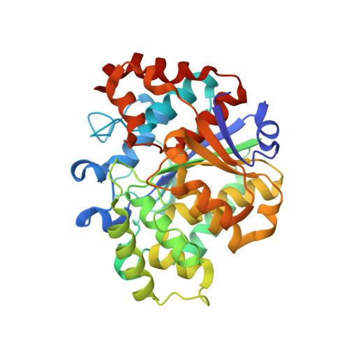
    Cobamides (Cbas) are coenzymes used by cells across all domains of life, but de novo synthesis is only found in some bacteria and archaea. Five enzymes assemble the nucleotide loop in the alpha phase of the corrin ring. Condensation of the activated ring and nucleobase yields adenosyl-Cba 5'-phosphate, which upon dephosphorylation yields the biologically active coenzyme (AdoCba). Base activation is catalyzed by a phosphoribosyltransferase (PRTase). The structure of the Salmonella enterica PRTase enzyme (i.e., SeCobT) is well-characterized, but archaeal PRTases are not. To gain insights into the mechanism of base activation by the PRTase from Methanocaldococcus jannaschii (MjCobT), we solved crystal structures of the enzyme in complex with substrate and products. We determined several structures: (i) a 2.2 Å structure of MjCobT in the absence of ligand (apo), (ii) structures of MjCobT bound to nicotinate mononucleotide (NaMN) and α-ribazole 5'-phosphate (α-RP) or α-adenylyl-5'-phosphate (α-AMP) at 2.3 and 1.4 Å, respectively. In MjCobT the general base that triggers the reaction is an aspartate residue (Asp 52) rather than a glutamate residue (E317) as in SeCobT. Notably, the dimer interface in MjCobT is completely different from that observed in SeCobT. Finally, entry PDB 3L0Z does not reflect the correct structure of MjCobT.


  • Organizational Affiliation
    • Department of Microbiology, University of Georgia, Athens, GA, 30602, USA.

Macromolecule Content 

  • Total Structure Weight: 76.51 kDa 
  • Atom Count: 5,325 
  • Modeled Residue Count: 687 
  • Deposited Residue Count: 700 
  • Unique protein chains: 1

Macromolecules

Find similar proteins by:|  3D Structure
Entity ID: 1
MoleculeChains  Sequence LengthOrganismDetailsImage
UPF0284 protein MJ1598
A, B
350Methanocaldococcus jannaschii DSM 2661Mutation(s): 0 
Gene Names: MJ1598
UniProt
Find proteins for Q58993 (Methanocaldococcus jannaschii (strain ATCC 43067 / DSM 2661 / JAL-1 / JCM 10045 / NBRC 100440))
Explore Q58993 
Go to UniProtKB:  Q58993
Entity Groups
Sequence Clusters30% Identity50% Identity70% Identity90% Identity95% Identity100% Identity
UniProt GroupQ58993
Sequence Annotations
Expand
Reference Sequence

Experimental Data & Validation

Experimental Data

  • Method: X-RAY DIFFRACTION
  • Resolution: 2.29 Å
  • R-Value Free:  0.240 (Depositor), 0.241 (DCC) 
  • R-Value Work:  0.193 (Depositor), 0.196 (DCC) 
  • R-Value Observed: 0.196 (Depositor) 
Space Group: P 1 21 1
Unit Cell:
Length ( Å )Angle ( ˚ )
a = 47.58α = 90
b = 139β = 111.67
c = 54.49γ = 90
Software Package:
Software NamePurpose
PHENIXrefinement
XDSdata reduction
XSCALEdata scaling
PHASERphasing
Cootmodel building

Structure Validation

View Full Validation Report


Ligand Structure Quality Assessment 


Entry History 

& Funding Information

Deposition Data


Funding OrganizationLocationGrant Number
National Institutes of Health/National Institute of General Medical Sciences (NIH/NIGMS)United StatesR35-GM130399

Revision History  (Full details and data files)

  • Version 1.0: 2020-07-22
    Type: Initial release
  • Version 1.1: 2023-10-11
    Changes: Data collection, Database references, Refinement description
  • Version 1.2: 2024-04-10
    Changes: Database references