6P55 | pdb_00006p55

Crystal structure of transpeptidase domain of PBP2 from Neisseria gonorrhoeae acylated by cefixime


Experimental Data Snapshot

  • Method: X-RAY DIFFRACTION
  • Resolution: 1.74 Å
  • R-Value Free: 
    0.188 (Depositor), 0.194 (DCC) 
  • R-Value Work: 
    0.160 (Depositor), 0.169 (DCC) 
  • R-Value Observed: 
    0.161 (Depositor) 

wwPDB Validation 3D Report Full Report

Validation slider image for 6P55

Ligand Structure Quality Assessment 


This is version 1.4 of the entry. See complete history

Literature

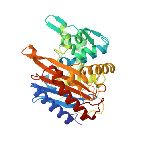
Recognition of the beta-lactam carboxylate triggers acylation ofNeisseria gonorrhoeaepenicillin-binding protein 2.

Singh, A.Tomberg, J.Nicholas, R.A.Davies, C.

(2019) J Biological Chem 294: 14020-14032

  • DOI: https://doi.org/10.1074/jbc.RA119.009942
  • Primary Citation Related Structures: 
    6P52, 6P53, 6P54, 6P55, 6P56

  • PubMed Abstract: 

    Resistance of Neisseria gonorrhoeae to extended-spectrum cephalosporins (ESCs) has become a major threat to human health. The primary mechanism by which N. gonorrhoeae becomes resistant to ESCs is by acquiring a mosaic penA allele, encoding penicillin-binding protein 2 (PBP2) variants containing up to 62 mutations compared with WT, of which a subset contribute to resistance. To interpret molecular mechanisms underpinning cephalosporin resistance, it is necessary to know how PBP2 is acylated by ESCs. Here, we report the crystal structures of the transpeptidase domain of WT PBP2 in complex with cefixime and ceftriaxone, along with structures of PBP2 in the apo form and with a phosphate ion bound in the active site at resolutions of 1-7-1.9 Å. These structures reveal that acylation of PBP2 by ESCs is accompanied by rotation of the Thr-498 side chain in the KTG motif to contact the cephalosporin carboxylate, twisting of the β3 strand to form the oxyanion hole, and rolling of the β3-β4 loop toward the active site. Recognition of the cephalosporin carboxylate appears to be the key trigger for formation of an acylation-competent state of PBP2. The structures also begin to explain the impact of mutations implicated in ESC resistance. In particular, a G545S mutation may hinder twisting of β3 because its side chain hydroxyl forms a hydrogen bond with Thr-498. Overall, our data suggest that acylation is initiated by conformational changes elicited or trapped by binding of ESCs and that these movements are restricted by mutations associated with resistance against ESCs.


  • Organizational Affiliation
    • Department of Biochemistry and Molecular Biology, Medical University of South Carolina, Charleston, South Carolina 29425.

Macromolecule Content 

  • Total Structure Weight: 72.17 kDa 
  • Atom Count: 5,462 
  • Modeled Residue Count: 653 
  • Deposited Residue Count: 658 
  • Unique protein chains: 1

Macromolecules

Find similar proteins by:|  3D Structure
Entity ID: 1
MoleculeChains  Sequence LengthOrganismDetailsImage
peptidoglycan D,D-transpeptidase PenA
A, B
329Neisseria gonorrhoeaeMutation(s): 0 
Gene Names: penA
EC: 3.4.16.4
UniProt
Find proteins for P08149 (Neisseria gonorrhoeae)
Explore P08149 
Go to UniProtKB:  P08149
Entity Groups
Sequence Clusters30% Identity50% Identity70% Identity90% Identity95% Identity100% Identity
UniProt GroupP08149
Sequence Annotations
Expand
Reference Sequence

Small Molecules

Ligands 3 Unique
IDChains Name / Formula / InChI Key2D Diagram3D Interactions
NZM
(Subject of Investigation/LOI)

Query on NZM


Download:Ideal Coordinates CCD File
C [auth A],
G [auth B]
cefixime, bound form
C16 H17 N5 O7 S2
LIFBJXSMIMSLKH-VSOMHRBSSA-N
PEG

Query on PEG


Download:Ideal Coordinates CCD File
F [auth A],
I [auth B],
J [auth B]
DI(HYDROXYETHYL)ETHER
C4 H10 O3
MTHSVFCYNBDYFN-UHFFFAOYSA-N
PO4

Query on PO4


Download:Ideal Coordinates CCD File
D [auth A],
E [auth A],
H [auth B]
PHOSPHATE ION
O4 P
NBIIXXVUZAFLBC-UHFFFAOYSA-K

Experimental Data & Validation

Experimental Data

  • Method: X-RAY DIFFRACTION
  • Resolution: 1.74 Å
  • R-Value Free:  0.188 (Depositor), 0.194 (DCC) 
  • R-Value Work:  0.160 (Depositor), 0.169 (DCC) 
  • R-Value Observed: 0.161 (Depositor) 
Space Group: P 1 21 1
Unit Cell:
Length ( Å )Angle ( ˚ )
a = 44.493α = 90
b = 78.013β = 90.54
c = 86.902γ = 90
Software Package:
Software NamePurpose
REFMACrefinement
HKL-2000data reduction
HKL-2000data scaling
FFTphasing

Structure Validation

View Full Validation Report


Ligand Structure Quality Assessment 


Entry History 

& Funding Information

Deposition Data


Funding OrganizationLocationGrant Number
National Institutes of Health/National Institute of General Medical Sciences (NIH/NIGMS)United StatesGM066861

Revision History  (Full details and data files)

  • Version 1.0: 2019-08-07
    Type: Initial release
  • Version 1.1: 2019-08-14
    Changes: Data collection, Database references
  • Version 1.2: 2019-10-02
    Changes: Data collection, Database references
  • Version 1.3: 2020-01-01
    Changes: Author supporting evidence
  • Version 1.4: 2024-11-13
    Changes: Data collection, Database references, Structure summary