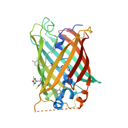
6OG8

Crystal structure of Green Fluorescent Protein (GFP); S65T, H148D with globally incorporated 3-F1Y; circular permutant (50-51)


Experimental Data Snapshot

  • Method: X-RAY DIFFRACTION
  • Resolution: 1.60 Å
  • R-Value Free: 0.190 
  • R-Value Work: 0.159 
  • R-Value Observed: 0.160 

Starting Model: experimental
View more details

wwPDB Validation   3D Report Full Report


This is version 2.1 of the entry. See complete history


Literature

Unusual Spectroscopic and Electric Field Sensitivity of Chromophores with Short Hydrogen Bonds: GFP and PYP as Model Systems.

Lin, C.Y.Boxer, S.G.

(2020) J Phys Chem B 124: 9513-9525

  • DOI: https://doi.org/10.1021/acs.jpcb.0c07730
  • Primary Citation of Related Structures:  
    6OG8, 6OG9, 6OGA, 6OGB, 6OGC, 6UN5, 6UN6, 6UN7

  • PubMed Abstract: 

    Short hydrogen bonds, with heavy-atom distances less than 2.7 Å, are believed to exhibit proton delocalization, and their possible role in catalysis has been widely debated. While spectroscopic and/or structural methods are usually employed to study the degree of proton delocalization, ambiguities still arise, and no direct information on the corresponding potential energy surface is obtained. Here, we apply an external electric field to perturb the short hydrogen bond(s) within a collection of green fluorescent protein S65T/H148D variants and photoactive yellow protein mutants, where the chromophore participates in the short hydrogen bond(s) and serves as an optical probe of the proton position. As the proton is charged, its position may shift in response to the external electric field, and the chromophore's electronic absorption can thus reflect the ease of proton transfer. The results suggest that low-barrier hydrogen bonds (LBHBs) are not present within these proteins even when proton affinities between donor and acceptor are closely matched. Exploiting the chromophores as precalibrated electrostatic probes, the covalency of short hydrogen bonds as a nonelectrostatic component is also revealed. A theoretical framework is developed to address a possible contribution of unusually large polarizabilities of short hydrogen bonds due to proton delocalization, but no clear evidence for this phenomenon is found in accordance with the absence of LBHBs.


  • Organizational Affiliation

    Department of Chemistry, Stanford University, Stanford, California 94305, United States.


Macromolecules
Find similar proteins by:  (by identity cutoff)  |  3D Structure
Entity ID: 1
MoleculeChains Sequence LengthOrganismDetailsImage
Crystal structure of Green Fluorescent Protein (GFP); S65T, H148D with globally incorporated 3-F1Y; circular permutant (50-51)A [auth B],
B [auth A]
251Aequorea victoriaMutation(s): 0 
UniProt
Find proteins for P42212 (Aequorea victoria)
Explore P42212 
Go to UniProtKB:  P42212
Entity Groups  
Sequence Clusters30% Identity50% Identity70% Identity90% Identity95% Identity100% Identity
UniProt GroupP42212
Sequence Annotations
Expand
  • Reference Sequence
Small Molecules
Modified Residues  2 Unique
IDChains TypeFormula2D DiagramParent
MFC
Query on MFC
A [auth B],
B [auth A]
L-PEPTIDE LINKINGC15 H16 F N3 O5GLY, TYR, GLY
YOF
Query on YOF
A [auth B],
B [auth A]
L-PEPTIDE LINKINGC9 H10 F N O3TYR
Experimental Data & Validation

Experimental Data

  • Method: X-RAY DIFFRACTION
  • Resolution: 1.60 Å
  • R-Value Free: 0.190 
  • R-Value Work: 0.159 
  • R-Value Observed: 0.160 
  • Space Group: P 1 21 1
Unit Cell:
Length ( Å )Angle ( ˚ )
a = 50.073α = 90
b = 70.072β = 95.93
c = 61.072γ = 90
Software Package:
Software NamePurpose
PHENIXrefinement
XDSdata reduction
XDSdata scaling
PHENIXphasing

Structure Validation

View Full Validation Report


Entry History & Funding Information

Deposition Data


Funding OrganizationLocationGrant Number
National Institutes of Health/National Institute of General Medical Sciences (NIH/NIGMS)United States118044

Revision History  (Full details and data files)

  • Version 1.0: 2020-04-08
    Type: Initial release
  • Version 1.1: 2020-10-28
    Changes: Database references
  • Version 1.2: 2020-11-04
    Changes: Database references
  • Version 1.3: 2023-10-11
    Changes: Data collection, Database references, Refinement description
  • Version 2.0: 2023-11-15
    Changes: Atomic model, Data collection, Derived calculations
  • Version 2.1: 2024-11-06
    Changes: Structure summary