6O9E | pdb_00006o9e

Structure of HIV-1 Reverse Transcriptase in complex with DNA and INDOPY-1


Experimental Data Snapshot

  • Method: X-RAY DIFFRACTION
  • Resolution: 2.40 Å
  • R-Value Free: 
    0.242 (Depositor), 0.249 (DCC) 
  • R-Value Work: 
    0.203 (Depositor), 0.210 (DCC) 
  • R-Value Observed: 
    0.204 (Depositor) 

Starting Model: experimental
View more details

wwPDB Validation 3D Report Full Report

Validation slider image for 6O9E

Ligand Structure Quality Assessment 


This is version 2.1 of the entry. See complete history

Literature

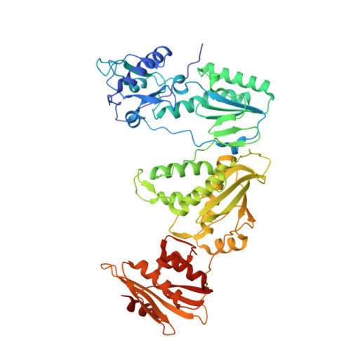
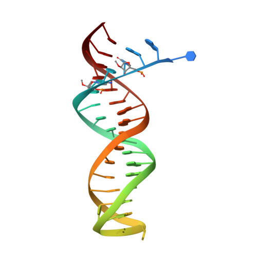
Structural Basis of HIV-1 Inhibition by Nucleotide-Competing Reverse Transcriptase Inhibitor INDOPY-1.

Ruiz, F.X.Hoang, A.Das, K.Arnold, E.

(2019) J Med Chem 62: 9996-10002

  • DOI: https://doi.org/10.1021/acs.jmedchem.9b01289
  • Primary Citation Related Structures: 
    6O9E

  • PubMed Abstract: 

    HIV-1 reverse transcriptase (RT) is an essential enzyme, targeting half of approved anti-AIDS drugs. While nucleoside RT inhibitors (NRTIs) are DNA chain terminators, the nucleotide-competing RT inhibitor (NcRTI) INDOPY-1 blocks dNTP binding to RT. Lack of structural information hindered INDOPY-1 improvement. Here we report the HIV-1 RT/DNA/INDOPY-1 crystal structure, revealing a unique mode of inhibitor binding at the polymerase active site without involving catalytic metal ions. The structure may enable new strategies for developing NcRTIs.


  • Organizational Affiliation
    • Rega Institute for Medical Research , 3000 Leuven , Belgium.

Macromolecule Content 

  • Total Structure Weight: 254.1 kDa 
  • Atom Count: 17,583 
  • Modeled Residue Count: 2,001 
  • Deposited Residue Count: 2,044 
  • Unique protein chains: 2
  • Unique nucleic acid chains: 1

Macromolecules


Find similar proteins by:|  3D Structure
Entity ID: 1
MoleculeChains  Sequence LengthOrganismDetailsImage
Reverse transcriptase p66
A, C
555Human immunodeficiency virus 1Mutation(s): 2 
EC: 2.7.7.49 (PDB Primary Data), 2.7.7.7 (PDB Primary Data), 3.1.26.13 (PDB Primary Data)
UniProt
Find proteins for P03366 (Human immunodeficiency virus type 1 group M subtype B (isolate BH10))
Explore P03366 
Go to UniProtKB:  P03366
Entity Groups
Sequence Clusters30% Identity50% Identity70% Identity90% Identity95% Identity100% Identity
UniProt GroupP03366
Sequence Annotations
Expand
Reference Sequence
Find similar proteins by:|  3D Structure
Entity ID: 2
MoleculeChains  Sequence LengthOrganismDetailsImage
Reverse transcriptase p51
B, D
429Human immunodeficiency virus 1Mutation(s): 1 
EC: 3.4.23.16
UniProt
Find proteins for P03366 (Human immunodeficiency virus type 1 group M subtype B (isolate BH10))
Explore P03366 
Go to UniProtKB:  P03366
Entity Groups
Sequence Clusters30% Identity50% Identity70% Identity90% Identity95% Identity100% Identity
UniProt GroupP03366
Sequence Annotations
Expand
Reference Sequence
Find similar nucleic acids by:  (by identity cutoff) 
Entity ID: 3
MoleculeChains LengthOrganismImage
DNA (38-MER)E [auth F],
F [auth E]
38synthetic construct
Sequence Annotations
Expand
Reference Sequence

Oligosaccharides

Help  
Entity ID: 4
MoleculeChains Length2D Diagram GlycosylationD Interactions
beta-D-fructofuranose-(2-1)-alpha-D-glucopyranose
G, H
2N/A
Glycosylation Resources
GlyTouCan: G05551OP
GlyCosmos: G05551OP

Small Molecules

Ligands 6 Unique
IDChains Name / Formula / InChI Key2D Diagram3D Interactions
IY1
(Subject of Investigation/LOI)

Query on IY1


Download:Ideal Coordinates CCD File
V [auth F],
X [auth E]
5-methyl-1-(4-nitrophenyl)-2-oxo-2,5-dihydro-1H-pyrido[3,2-b]indole-3-carbonitrile
C19 H12 N4 O3
TZEXGCXFOXXFKH-UHFFFAOYSA-N
PG4

Query on PG4


Download:Ideal Coordinates CCD File
W [auth E]TETRAETHYLENE GLYCOL
C8 H18 O5
UWHCKJMYHZGTIT-UHFFFAOYSA-N
PEG

Query on PEG


Download:Ideal Coordinates CCD File
P [auth B],
Q [auth B]
DI(HYDROXYETHYL)ETHER
C4 H10 O3
MTHSVFCYNBDYFN-UHFFFAOYSA-N
SO4

Query on SO4


Download:Ideal Coordinates CCD File
I [auth A]
J [auth A]
L [auth B]
M [auth B]
R [auth C]
I [auth A],
J [auth A],
L [auth B],
M [auth B],
R [auth C],
S [auth C]
SULFATE ION
O4 S
QAOWNCQODCNURD-UHFFFAOYSA-L
GOL

Query on GOL


Download:Ideal Coordinates CCD File
N [auth B],
O [auth B],
T [auth C]
GLYCEROL
C3 H8 O3
PEDCQBHIVMGVHV-UHFFFAOYSA-N
NH4

Query on NH4


Download:Ideal Coordinates CCD File
K [auth A],
U [auth C]
AMMONIUM ION
H4 N
QGZKDVFQNNGYKY-UHFFFAOYSA-O
Binding Affinity Annotations 
IDSourceBinding Affinity
IY1 BindingDB:  6O9E IC50: min: 19.7, max: 1123 (nM) from 3 assay(s)

Biologically Interesting Molecules (External Reference) 

1 Unique

Experimental Data & Validation

Experimental Data

  • Method: X-RAY DIFFRACTION
  • Resolution: 2.40 Å
  • R-Value Free:  0.242 (Depositor), 0.249 (DCC) 
  • R-Value Work:  0.203 (Depositor), 0.210 (DCC) 
  • R-Value Observed: 0.204 (Depositor) 
Space Group: P 1 21 1
Unit Cell:
Length ( Å )Angle ( ˚ )
a = 89.762α = 90
b = 127.663β = 101.5
c = 131.61γ = 90
Software Package:
Software NamePurpose
PHENIXrefinement
MOSFLMdata reduction
Aimlessdata scaling
PHASERphasing

Structure Validation

View Full Validation Report


Ligand Structure Quality Assessment 


Entry History 

& Funding Information

Deposition Data


Funding OrganizationLocationGrant Number
National Institutes of Health/National Institute of General Medical Sciences (NIH/NIGMS)United StatesMERIT Award R37 AI027690

Revision History  (Full details and data files)

  • Version 1.0: 2019-10-23
    Type: Initial release
  • Version 1.1: 2019-11-06
    Changes: Data collection, Database references
  • Version 1.2: 2019-11-27
    Changes: Database references
  • Version 1.3: 2020-01-01
    Changes: Author supporting evidence
  • Version 2.0: 2020-07-29
    Type: Remediation
    Reason: Carbohydrate remediation
    Changes: Advisory, Atomic model, Data collection, Derived calculations, Non-polymer description, Structure summary
  • Version 2.1: 2023-10-11
    Changes: Data collection, Database references, Refinement description, Structure summary