6MOE | pdb_00006moe

Monomeric DARPin E2 complex with EpoR


Experimental Data Snapshot

  • Method: X-RAY DIFFRACTION
  • Resolution: 2.09 Å
  • R-Value Free: 
    0.230 (Depositor), 0.234 (DCC) 
  • R-Value Work: 
    0.204 (Depositor), 0.206 (DCC) 
  • R-Value Observed: 
    0.205 (Depositor) 

Starting Model: experimental
View more details

wwPDB Validation 3D Report Full Report

Validation slider image for 6MOE

This is version 1.3 of the entry. See complete history

Literature

Topological control of cytokine receptor signaling induces differential effects in hematopoiesis.

Mohan, K.Ueda, G.Kim, A.R.Jude, K.M.Fallas, J.A.Guo, Y.Hafer, M.Miao, Y.Saxton, R.A.Piehler, J.Sankaran, V.G.Baker, D.Garcia, K.C.

(2019) Science 364

  • DOI: https://doi.org/10.1126/science.aav7532
  • Primary Citation Related Structures: 
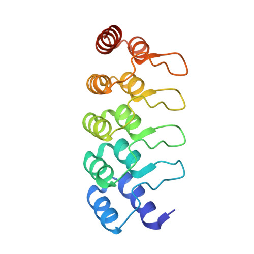
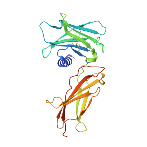
    6MOE, 6MOF, 6MOG, 6MOH, 6MOI, 6MOJ, 6MOK, 6MOL

  • PubMed Abstract: 

    Although tunable signaling by G protein-coupled receptors can be exploited through medicinal chemistry, a comparable pharmacological approach has been lacking for the modulation of signaling through dimeric receptors, such as those for cytokines. We present a strategy to modulate cytokine receptor signaling output by use of a series of designed C2-symmetric cytokine mimetics, based on the designed ankyrin repeat protein (DARPin) scaffold, that can systematically control erythropoietin receptor (EpoR) dimerization orientation and distance between monomers. We sampled a range of EpoR geometries by varying intermonomer angle and distance, corroborated by several ligand-EpoR complex crystal structures. Across the range, we observed full, partial, and biased agonism as well as stage-selective effects on hematopoiesis. This surrogate ligand strategy opens access to pharmacological modulation of therapeutically important cytokine and growth factor receptor systems.


  • Organizational Affiliation
    • Department of Molecular and Cellular Physiology, Stanford University School of Medicine, Stanford, CA 94305, USA.

Macromolecule Content 

  • Total Structure Weight: 87.41 kDa 
  • Atom Count: 6,249 
  • Modeled Residue Count: 755 
  • Deposited Residue Count: 794 
  • Unique protein chains: 2

Macromolecules

Find similar proteins by:|  3D Structure
Entity ID: 1
MoleculeChains  Sequence LengthOrganismDetailsImage
E2 DARPin
A, B
168synthetic constructMutation(s): 0 
Find similar proteins by:|  3D Structure
Entity ID: 2
MoleculeChains  Sequence LengthOrganismDetailsImage
Erythropoietin receptor
C, D
229Homo sapiensMutation(s): 0 
Gene Names: EPOR
UniProt & NIH Common Fund Data Resources
Find proteins for P19235 (Homo sapiens)
Explore P19235 
Go to UniProtKB:  P19235
PHAROS:  P19235
GTEx:  ENSG00000187266 
Entity Groups
Sequence Clusters30% Identity50% Identity70% Identity90% Identity95% Identity100% Identity
UniProt GroupP19235
Sequence Annotations
Expand
Reference Sequence

Small Molecules

Ligands 3 Unique
IDChains Name / Formula / InChI Key2D Diagram3D Interactions
EPE

Query on EPE


Download:Ideal Coordinates CCD File
I [auth B]4-(2-HYDROXYETHYL)-1-PIPERAZINE ETHANESULFONIC ACID
C8 H18 N2 O4 S
JKMHFZQWWAIEOD-UHFFFAOYSA-N
PO4

Query on PO4


Download:Ideal Coordinates CCD File
E [auth A]
F [auth A]
H [auth B]
J [auth C]
K [auth C]
E [auth A],
F [auth A],
H [auth B],
J [auth C],
K [auth C],
L [auth C],
N [auth D],
O [auth D],
P [auth D]
PHOSPHATE ION
O4 P
NBIIXXVUZAFLBC-UHFFFAOYSA-K
EDO

Query on EDO


Download:Ideal Coordinates CCD File
G [auth A],
M [auth C]
1,2-ETHANEDIOL
C2 H6 O2
LYCAIKOWRPUZTN-UHFFFAOYSA-N

Experimental Data & Validation

Experimental Data

  • Method: X-RAY DIFFRACTION
  • Resolution: 2.09 Å
  • R-Value Free:  0.230 (Depositor), 0.234 (DCC) 
  • R-Value Work:  0.204 (Depositor), 0.206 (DCC) 
  • R-Value Observed: 0.205 (Depositor) 
Space Group: P 21 21 21
Unit Cell:
Length ( Å )Angle ( ˚ )
a = 69.25α = 90
b = 122.16β = 90
c = 132.57γ = 90
Software Package:
Software NamePurpose
PHENIXrefinement
XDSdata reduction
XDSdata scaling
PHASERphasing

Structure Validation

View Full Validation Report


Entry History 

Deposition Data

Revision History  (Full details and data files)

  • Version 1.0: 2019-06-05
    Type: Initial release
  • Version 1.1: 2019-06-12
    Changes: Data collection, Database references
  • Version 1.2: 2023-10-11
    Changes: Data collection, Database references, Refinement description
  • Version 1.3: 2024-10-23
    Changes: Structure summary