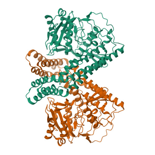
6IRV

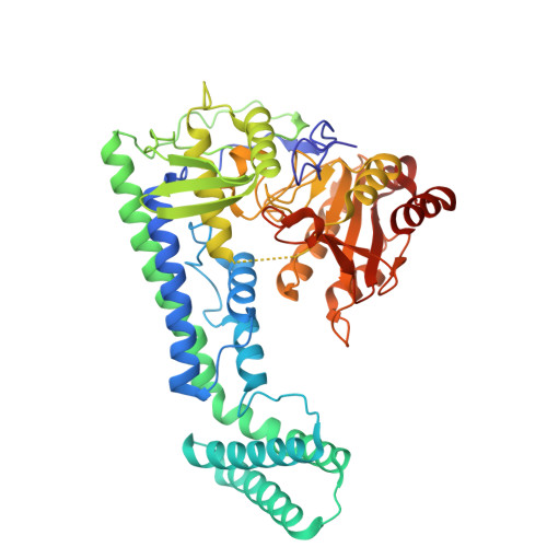
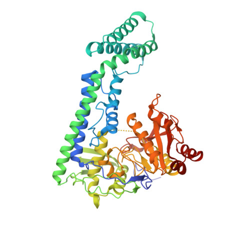
Crystal structure of the human cap-specific adenosine methyltransferase


Experimental Data Snapshot

  • Method: X-RAY DIFFRACTION
  • Resolution: 2.70 Å
  • R-Value Free: 0.252 
  • R-Value Work: 0.214 
  • R-Value Observed: 0.216 

Starting Model: experimental
View more details

wwPDB Validation   3D Report Full Report


This is version 1.3 of the entry. See complete history


Literature

Cap-specific terminal N 6 -methylation of RNA by an RNA polymerase II-associated methyltransferase.

Akichika, S.Hirano, S.Shichino, Y.Suzuki, T.Nishimasu, H.Ishitani, R.Sugita, A.Hirose, Y.Iwasaki, S.Nureki, O.Suzuki, T.

(2019) Science 363

  • DOI: https://doi.org/10.1126/science.aav0080
  • Primary Citation of Related Structures:  
    6IRV, 6IRW, 6IRX, 6IRY, 6IRZ, 6IS0

  • PubMed Abstract: 

    N 6 -methyladenosine (m 6 A), a major modification of messenger RNAs (mRNAs), plays critical roles in RNA metabolism and function. In addition to the internal m 6 A, N 6 , 2'- O -dimethyladenosine (m 6 Am) is present at the transcription start nucleotide of capped mRNAs in vertebrates. However, its biogenesis and functional role remain elusive. Using a reverse genetics approach, we identified PCIF1, a factor that interacts with the serine-5-phosphorylated carboxyl-terminal domain of RNA polymerase II, as a cap-specific adenosine methyltransferase (CAPAM) responsible for N 6 -methylation of m 6 Am. The crystal structure of CAPAM in complex with substrates revealed the molecular basis of cap-specific m 6 A formation. A transcriptome-wide analysis revealed that N 6 -methylation of m 6 Am promotes the translation of capped mRNAs. Thus, a cap-specific m 6 A writer promotes translation of mRNAs starting from m 6 Am.


  • Organizational Affiliation

    Department of Chemistry and Biotechnology, Graduate School of Engineering, The University of Tokyo, 7-3-1 Hongo, Bunkyo-ku, Tokyo 113-8656, Japan.


Macromolecules
Find similar proteins by:  (by identity cutoff)  |  3D Structure
Entity ID: 1
MoleculeChains Sequence LengthOrganismDetailsImage
Phosphorylated CTD-interacting factor 1
A, B
508Homo sapiensMutation(s): 0 
Gene Names: PCIF1C20orf67
EC: 2.1.1.62
UniProt & NIH Common Fund Data Resources
Find proteins for Q9H4Z3 (Homo sapiens)
Explore Q9H4Z3 
Go to UniProtKB:  Q9H4Z3
PHAROS:  Q9H4Z3
GTEx:  ENSG00000100982 
Entity Groups  
Sequence Clusters30% Identity50% Identity70% Identity90% Identity95% Identity100% Identity
UniProt GroupQ9H4Z3
Sequence Annotations
Expand
  • Reference Sequence
Experimental Data & Validation

Experimental Data

  • Method: X-RAY DIFFRACTION
  • Resolution: 2.70 Å
  • R-Value Free: 0.252 
  • R-Value Work: 0.214 
  • R-Value Observed: 0.216 
  • Space Group: P 21 21 21
Unit Cell:
Length ( Å )Angle ( ˚ )
a = 70.699α = 90
b = 120.309β = 90
c = 156.831γ = 90
Software Package:
Software NamePurpose
Aimlessdata scaling
PHENIXrefinement
PDB_EXTRACTdata extraction
XDSdata reduction
MOLREPphasing

Structure Validation

View Full Validation Report


Entry History 

Deposition Data

Revision History  (Full details and data files)

  • Version 1.0: 2018-12-05
    Type: Initial release
  • Version 1.1: 2019-01-23
    Changes: Data collection, Database references
  • Version 1.2: 2020-10-28
    Changes: Database references
  • Version 1.3: 2023-11-22
    Changes: Data collection, Database references, Refinement description