Structure-function relationships underlying the dualN-acetylmuramic andN-acetylglucosamine specificities of the bacterial peptidoglycan deacetylase PdaC.
Grifoll-Romero, L., Sainz-Polo, M.A., Albesa-Jove, D., Guerin, M.E., Biarnes, X., Planas, A.(2019) J Biological Chem 294: 19066-19080
- PubMed: 31690626 Search on PubMedSearch on PubMed Central
- DOI: https://doi.org/10.1074/jbc.RA119.009510
- Primary Citation Related Structures:
6H8L, 6H8N - PubMed Abstract:
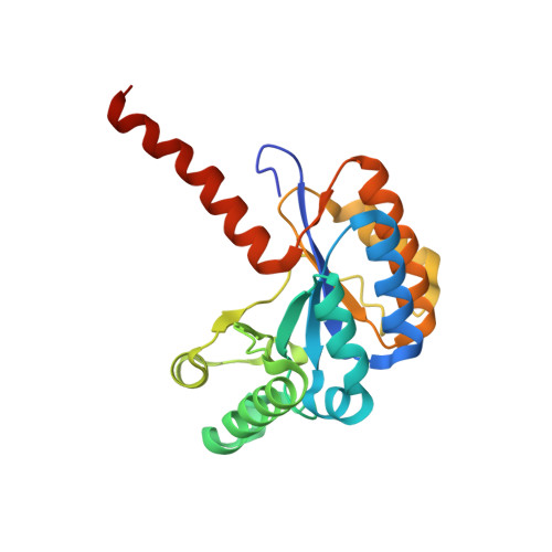
Bacillus subtilis PdaC ( Bs PdaC) is a membrane-bound, multidomain peptidoglycan N- deacetylase acting on N -acetylmuramic acid (MurNAc) residues and conferring lysozyme resistance to modified cell wall peptidoglycans. Bs PdaC contains a C-terminal family 4 carbohydrate esterase (CE4) catalytic domain, but unlike other MurNAc deacetylases, Bs PdaC also has GlcNAc deacetylase activity on chitooligosaccharides (COSs), characteristic of chitin deacetylases. To uncover the molecular basis of this dual activity, here we determined the X-ray structure of the Bs PdaC CE4 domain at 1.54 Å resolution and analyzed its mode of action on COS substrates. We found that the minimal substrate is GlcNAc 3 and that activity increases with the degree of glycan polymerization. COS deacetylation kinetics revealed that Bs PdaC operates by a multiple-chain mechanism starting at the internal GlcNAc units and leading to deacetylation of all but the reducing-end GlcNAc residues. Interestingly, Bs PdaC shares higher sequence similarity with the peptidoglycan GlcNAc deacetylase Sp PgdaA than with other MurNAc deacetylases. Therefore, we used ligand-docking simulations to analyze the dual GlcNAc- and MurNAc-binding specificities of Bs PdaC and compared them with those of Sp PgdA and Bs PdaA, representing peptidoglycan deacetylases highly specific for GlcNAc or MurNAc residues, respectively. Bs PdaC retains the conserved Asp-His-His metal-binding triad characteristic of CE4 enzymes acting on GlcNAc residues, differing from MurNAc deacetylases that lack the metal-coordinating Asp residue. Bs PdaC contains short loops similar to those in Sp PgdA, resulting in an open binding cleft that can accommodate polymeric substrates. We propose that PdaC is the first member of a new subclass of peptidoglycan MurNAc deacetylases.
- Laboratory of Biochemistry, Institut Químic de Sarrià, University Ramon Llull, 08017 Barcelona, Spain.
Organizational Affiliation: