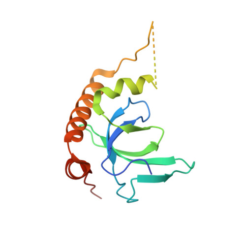
6G2O

X-ray structure of NSD3-PWWP1 in complex with compound BI-9321


Experimental Data Snapshot

  • Method: X-RAY DIFFRACTION
  • Resolution: 1.81 Å
  • R-Value Free: 0.224 
  • R-Value Work: 0.193 
  • R-Value Observed: 0.195 

wwPDB Validation   3D Report Full Report


Ligand Structure Quality Assessment 


This is version 1.2 of the entry. See complete history


Literature

Fragment-based discovery of a chemical probe for the PWWP1 domain of NSD3.

Bottcher, J.Dilworth, D.Reiser, U.Neumuller, R.A.Schleicher, M.Petronczki, M.Zeeb, M.Mischerikow, N.Allali-Hassani, A.Szewczyk, M.M.Li, F.Kennedy, S.Vedadi, M.Barsyte-Lovejoy, D.Brown, P.J.Huber, K.V.M.Rogers, C.M.Wells, C.I.Fedorov, O.Rumpel, K.Zoephel, A.Mayer, M.Wunberg, T.Bose, D.Zahn, S.Arnhof, H.Berger, H.Reiser, C.Hormann, A.Krammer, T.Corcokovic, M.Sharps, B.Winkler, S.Haring, D.Cockcroft, X.L.Fuchs, J.E.Mullauer, B.Weiss-Puxbaum, A.Gerstberger, T.Boehmelt, G.Vakoc, C.R.Arrowsmith, C.H.Pearson, M.McConnell, D.B.

(2019) Nat Chem Biol 15: 822-829

  • DOI: https://doi.org/10.1038/s41589-019-0310-x
  • Primary Citation of Related Structures:  
    6G24, 6G25, 6G27, 6G29, 6G2B, 6G2C, 6G2E, 6G2F, 6G2O, 6G3P, 6G3T

  • PubMed Abstract: 

    Here, we report the fragment-based discovery of BI-9321, a potent, selective and cellular active antagonist of the NSD3-PWWP1 domain. The human NSD3 protein is encoded by the WHSC1L1 gene located in the 8p11-p12 amplicon, frequently amplified in breast and squamous lung cancer. Recently, it was demonstrated that the PWWP1 domain of NSD3 is required for the viability of acute myeloid leukemia cells. To further elucidate the relevance of NSD3 in cancer biology, we developed a chemical probe, BI-9321, targeting the methyl-lysine binding site of the PWWP1 domain with sub-micromolar in vitro activity and cellular target engagement at 1 µM. As a single agent, BI-9321 downregulates Myc messenger RNA expression and reduces proliferation in MOLM-13 cells. This first-in-class chemical probe BI-9321, together with the negative control BI-9466, will greatly facilitate the elucidation of the underexplored biological function of PWWP domains.


  • Organizational Affiliation

    Boehringer Ingelheim RCV GmbH & Co KG, Vienna, Austria. jark.boettcher@boehringer-ingelheim.com.


Macromolecules
Find similar proteins by:  (by identity cutoff)  |  3D Structure
Entity ID: 1
MoleculeChains Sequence LengthOrganismDetailsImage
Histone-lysine N-methyltransferase NSD3136Homo sapiensMutation(s): 0 
Gene Names: NSD3WHSC1L1DC28
EC: 2.1.1.43 (PDB Primary Data), 2.1.1.370 (UniProt), 2.1.1.371 (UniProt)
UniProt & NIH Common Fund Data Resources
Find proteins for Q9BZ95 (Homo sapiens)
Explore Q9BZ95 
Go to UniProtKB:  Q9BZ95
PHAROS:  Q9BZ95
GTEx:  ENSG00000147548 
Entity Groups  
Sequence Clusters30% Identity50% Identity70% Identity90% Identity95% Identity100% Identity
UniProt GroupQ9BZ95
Sequence Annotations
Expand
  • Reference Sequence
Small Molecules
Ligands 1 Unique
IDChains Name / Formula / InChI Key2D Diagram3D Interactions
EJE
Query on EJE

Download Ideal Coordinates CCD File 
B [auth A][4-[5-(7-fluoranylquinolin-4-yl)-1-methyl-imidazol-4-yl]-3,5-dimethyl-phenyl]methanamine
C22 H21 F N4
WOAOENGFAAUUGT-UHFFFAOYSA-N
Experimental Data & Validation

Experimental Data

  • Method: X-RAY DIFFRACTION
  • Resolution: 1.81 Å
  • R-Value Free: 0.224 
  • R-Value Work: 0.193 
  • R-Value Observed: 0.195 
  • Space Group: P 21 21 21
Unit Cell:
Length ( Å )Angle ( ˚ )
a = 44.279α = 90
b = 47.568β = 90
c = 61.113γ = 90
Software Package:
Software NamePurpose
XDSdata reduction
Aimlessdata scaling
BUSTERrefinement
PDB_EXTRACTdata extraction
BALBESphasing

Structure Validation

View Full Validation Report


Ligand Structure Quality Assessment 


Entry History 

Deposition Data

Revision History  (Full details and data files)

  • Version 1.0: 2019-06-26
    Type: Initial release
  • Version 1.1: 2020-01-15
    Changes: Database references
  • Version 1.2: 2024-05-08
    Changes: Data collection, Database references